Vital Sign Measurement Across the Lifespan - 2nd Canadian Edition by Jennifer L. Lapum, Margaret Verkuyl, Wendy Garcia, Oona St-Amant, and Andy Tan is licensed under a Creative Commons Attribution 4.0 International License, except where otherwise noted.
© 2021 Ryerson University
The CC licence permits you to retain, reuse, copy, redistribute, and revise this book—in whole or in part—for free providing the authors are attributed as follows:
More specific attribution information is provided at the end of each chapter section that includes adapted content.
The 2nd Canadian Edition of this textbook includes over 120 original H5P activities embedded throughout the text. These activities can be attributed as follows:
The 1st Canadian Edition can be accessed here: Vital Sign Measurement Across the Lifespan – 1st Canadian Edition
If you redistribute all or part of this book, it is recommended the following statement be added to the copyright page so readers can access the original book at no cost:
Sample APA-style citation:
This textbook can be referenced. In APA citation style (7th edition), it would appear as follows:
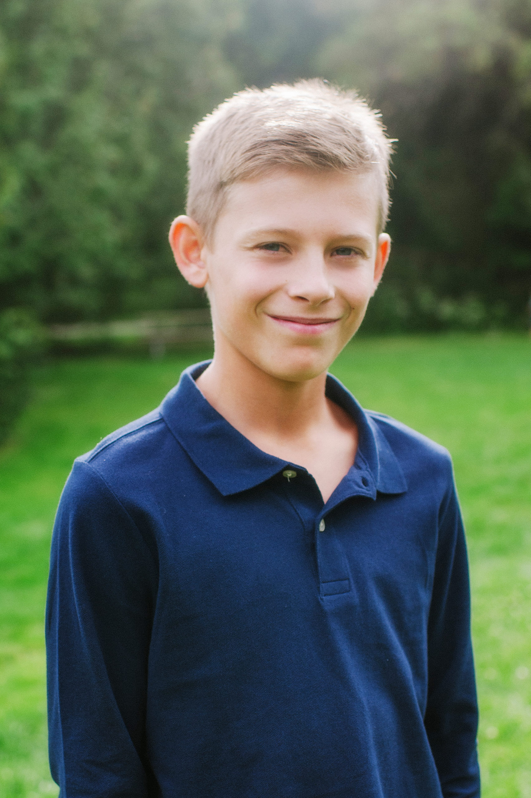
Cover image attribution:
Ebook ISBN: 978-1-77420-087-2
Print ISBN: 978-1-77420-086-5
Visit BCcampus Open Education to learn about open education in British Columbia.
This textbook is licensed under a Creative Commons Attribution 4.0 International (CC BY) license, which means that you are free to:
The licensor cannot revoke these freedoms as long as you follow the license terms.
The purpose of this textbook is to help learners develop best practices in vital sign measurement. Its intended audience is students in health-related post-secondary programs as well as healthcare providers. Using a multi-media and interactive approach, it will provide opportunities to read about, observe, practice, and test vital sign measurement.
Note that this book is best viewed via the online webbook on a computer or mobile device so that you can watch the videos and complete the interactive activities. However, it is also available in the following formats:
You can access the online webbook and download any of the formats for free here: Vital Sign Measurement Across the Lifespan – 2nd Canadian Edition. To download the book in a different format, look for the “Download this book” drop-down menu and select the file type you want.
This textbook will provide you with the opportunity to read about, observe, practice, and test vital sign measurement. Boxes with helpful tips are provided throughout the chapters:
A Chapter Summary and Printable Flashcards highlighting techniques for each vital sign measurement are provided at the end of each chapter. These printable flashcards are all located together in the textbook’s conclusion chapter.
You can review the full textbook or advance to sections that you have identified as areas you want to work on. The textbook has a self-directed format and provides an interactive and engaging way for you to learn about and develop competence in the measurement of vital signs while integrating knowledge about anatomy and physiology.
You will learn about various vital signs including temperature, pulse, respiration, blood pressure, and oxygen saturation. Measurement of vital signs is a foundational, psychomotor skill for healthcare providers and students in post-secondary health-related programs. These measurements provide information about a person’s overall state of health and more specifically about their cardiovascular and respiratory status. These measurements can also reveal changes in a client’s vital signs over time and changes in their overall state of health. Proficiency in vital sign measurement is essential to client safety, care, and management. Measurements can influence clinical decision-making related to therapeutic interventions.
An interactive or media element has been excluded from this version of the text. You can view it online here:
https://opentextbc.ca/vitalsignmeasurement/?p=331
Learning Outcomes
It is important to seek informed consent while creating a therapeutic and safe environment during all encounters with clients. You will usually begin by introducing yourself by name and designation so the client knows who you are. Next, explain what you are going to do and always ask permission to touch before beginning vital sign measurement. For example, an appropriate introduction is:
It is also important to ensure the client’s privacy by closing the curtains or the door to the room.
Clean hands and clean equipment are essential to infection prevention and control when measuring vital signs. Ensuring cleanliness helps reduce communicable and infectious diseases, particularly health care-associated infections, which are infectious organisms acquired by a client while in hospital. Common infections include clostridium difficile (C. diff), vancomycin-resistant enterococcus (VRE), and methicillin-resistant staphylococcus aureus (MRSA).
Ensuring your hands are clean is the best way to prevent and control infection. Hand hygiene can include cleansing with hand gel (see Figure 1.1) and hand washing. (see Figure 1.2). Use an alcohol-based sanitizer before and after contact with clients. Place gel on your hands and rub all hand surfaces for at least fifteen seconds. When washing hands using soap and water, wet your hands and apply soap. Rub all hand surfaces for about fifteen seconds, then rinse your hands. If the tap is not automatic, then turn it off with a paper towel.
Points to Consider
Hand gel is the preferred method of hand hygiene because it kills more bacteria and is easily accessible to healthcare providers. Soap and water is used when hands/gloves come into contact with bodily fluids.
While watching the interactive video on applying hand gel, apply your knowledge and critical thinking skills by answering the questions throughout the video. Please note: there is no sound in this video.
An interactive or media element has been excluded from this version of the text. You can view it online here:
https://opentextbc.ca/vitalsignmeasurement/?p=28
While watching the interactive video on hand washing, perform the skill along with the student while applying your knowledge and critical thinking skills to answer the questions throughout the video. Please note: there is no sound in this video.
An interactive or media element has been excluded from this version of the text. You can view it online here:
https://opentextbc.ca/vitalsignmeasurement/?p=28
Healthcare providers always inspect equipment before use to ensure it is in good working condition. Equipment (e.g., stethoscopes, pulse oximeters) can be cleaned with alcohol-based solutions to disinfect the surfaces. Automated devices should be regularly serviced to ensure accuracy. Biomedical technicians/experts are responsible for preventative maintenance and calibration to optimize functioning.
Click on the hotspots to learn about the following pieces of equipment:
An interactive or media element has been excluded from this version of the text. You can view it online here:
https://opentextbc.ca/vitalsignmeasurement/?p=28
A pain assessment is conducted in conjunction with the measurement of vital signs because pain can influence the findings. Pain can activate the sympathetic nervous system and increase pulse, respiration, and blood pressure. Pain is a complex issue, and a comprehensive discussion of pain assessment is beyond the scope of this e-book.
Briefly, because pain is subjective, self-reports are the most effective way to assess pain. The choice of pain assessment tool depends on the client situation: healthcare providers frequently use a numeric rating scale such as “rate your pain on a scale of 0 to 10 with zero being no pain and ten being the very worst pain that you have ever felt.” The response is often recorded on the vital sign record and expanded on in the narrative notes. Another common tool is the PQRSTU mneumonic in which each letter corresponds to a series of questions.
The order of vital sign measurement is influenced by the client situation. Healthcare providers often place the pulse oximeter probe on a client while proceeding to take pulse, respiration, blood pressure, and temperature. However, in some situations this order is modified and the healthcare provider needs to critically assess the situation to prioritize the vital sign measurement order. For example, with newborns/infants, it is best to proceed from least invasive to most invasive, so it is best to begin with respiration, pulse, oxygen saturation, temperature and if required, blood pressure. In an emergency situation or if a person loses consciousness, it is best to begin with pulse and blood pressure. Generally, it is important to conduct a complete set of vital signs unless otherwise indicated.
Determining the significance of vital sign measurements involves a process of diagnostic reasoning. The healthcare provider analyzes client data and makes decisions about whether the vital signs are normal or abnormal and whether the findings are significant: the following chapters provide normal vital sign ranges. The healthcare provider also considers agency policy, if applicable, about vital sign ranges to assess any abnormal variations and clinical significance. Additionally, the healthcare provider considers the client’s baseline vital signs to obtain a better sense of the client’s ‘normal’ and allow comparison (e.g., of trends) over time. The diagnostic reasoning process also involves considering other available objective and subjective data.
Timely documentation of vital sign measurements is imperative as a form of communication, to observe trends in vital sign measurements, and to ensure effective intervention when needed. Documentation occurs on paper-based vital sign records or electronic systems depending on the agency. Healthcare providers follow the agency’s documentation policy and the professional standards of practice. If using a vital sign record, healthcare providers use the symbols noted on the legend of the record.
Please answer the seven questions in the following question set.
An interactive or media element has been excluded from this version of the text. You can view it online here:
https://opentextbc.ca/vitalsignmeasurement/?p=28
Temperature refers to the degree of heat or cold in an object or a human body. In humans, the brain’s hypothalamus acts as the body’s thermostat and is responsible for regulating its temperature (OER #2). See Figure 2.1 of the hypothalamus.
The human body is constantly adapting to internal health states and environmental conditions, and the hypothalamus is programmed to tell the body to generate heat if the body temperature is low. For example, the hypothalamus can activate peripheral vasoconstriction and shivering (contraction of skeletal muscles) to prevent a decrease in body temperature. The hypothalamus can also reduce heat if the body temperature is too high. For example, it can activate peripheral vasodilation to increase heat loss and cause a person to perspire, which cools the body.
Part of this content was adapted from OER #2 (as noted in brackets above):
Healthcare providers measure a client’s temperature because it can give information about their state of health and influence clinical decisions. Accurate measurements and interpretation are vital so that hyperthermia and hypothermia can be identified and appropriate interventions determined.
Hyperthermia refers to an elevated body temperature. It can be related to an internal or external source. External sources that increase body temperature could include exposure to excessive heat on a hot day or being in a sauna or hot tub. Internal sources that may increase body temperature include fever caused by an infection or tissue breakdown associated with physical trauma (e.g., surgery, myocardial infarction) or some neurological conditions (e.g., cerebral vascular accident, cerebral edema, brain tumour). Hyperthermia that is associated with an infectious agent, such as a bacteria or virus (e.g., the flu) is referred to as febrile. Unresolved hyperthermic body states can lead to cell damage.
Hypothermia refers to a lowered body temperature. It is usually related to an external source such as being exposed to the cold for an extended period of time. Hypothermia is sometimes purposefully induced during surgery, or for certain medical conditions, to reduce the body’s need for oxygen. Unresolved hypothermic body states can slow cellular processes and lead to loss of consciousness.
An interactive or media element has been excluded from this version of the text. You can view it online here:
https://opentextbc.ca/vitalsignmeasurement/?p=34
Methods of measuring a client’s body temperature vary based on developmental age, cognitive functioning, level of consciousness, state of health, safety, and agency/unit policy. The healthcare provider chooses the best method after considering client safety, accuracy, and least invasiveness, all contingent on the client’s health and illness state. The most accurate way to measure core body temperature is an invasive method through a pulmonary artery catheter. This is only performed in a critical care area when constant measurements are required along with other life-saving interventions.
Methods of measurement include oral, axillary, tympanic, rectal, and dermal routes.
Oral temperature can be taken with clients who can follow instructions, so this kind of measurement is common for clients over the age of four, or even younger children if they are cooperative. Another route other than oral (e.g., tympanic or axillary) is preferable when a client is on oxygen delivered via a face mask because this can alter the temperature.
For children younger than four, axillary temperature is commonly measured unless a more accurate reading is required.
Rectal temperature is an accurate way to measure body temperature (Mazerolle, Ganio, Casa, Vingren, & Klau, 2011). The rectal route is recommended by the Canadian Pediatric Society for children under two years of age (Leduc & Woods, 2017). However, this method is not used on infants younger than thirty days or premature infants because of the risk of rectal tearing. If the rectal method is required, the procedure is generally only used by nurses and physicians.
Dermal routes are alternative methods of measurement that may be used in some agencies and practice areas. This method can involve holding the device and sliding it over the skin of the forehead and then down over the temporal artery in one motion. Dermal strips can also be placed on the forehead to measure skin temperature, but are not yet widely used, and the accuracy of this method has not yet been verified. More recently, there has been an increase in non-contact infrared thermometers particularly in the era of COVID-19 and other highly transmissible diseases. Depending on the type, these thermometers can be held at a short distance from the forehead or temporal area to measure temperature. Alternatively, some handheld thermal scanners that use an infrared camera can be held at a greater distance to screen large masses of people. Please refer to the manufacturer’s suggested reference range for non-contact infrared thermometers and thermal scanners.
While watching the interactive video on measuring temperature with a non-contact infrared thermometer, apply your knowledge and critical thinking skills to answer the questions throughout the video. Please note: there is no sound in this video.
An interactive or media element has been excluded from this version of the text. You can view it online here:
https://opentextbc.ca/vitalsignmeasurement/?p=36
Points to Consider
The accuracy of measurements is most often influenced by the healthcare provider’s adherence to the correct technique.
The following pages detail the normal temperature ranges and techniques associated with oral, axillary, tympanic, and rectal temperature methods.
The human body’s core temperature (internal body temperature) is measured in degrees Celsius (ºC) or Fahrenheit (ºF). In Canada, degrees Celsius is most commonly used.
In adults, the normal core body temperature (referred to as normothermia or afebrile) is 36.5–37.5ºC or 97.7–99.5ºF (OER #2).
A wider temperature range is acceptable in infants and young children, and can range from 35.5–37.7ºC or 95.9–99.8ºF. Infants and children have a wider temperature range because their heat control mechanisms are less effective. They are at risk for heat loss for many reasons including having less subcutaneous fat than adults, a larger body surface area in comparison to weight (and larger head size in proportion to the rest of the body), immature metabolic mechanisms (e.g., they may be unable to shiver), and limited ability to produce heat through activity. They are also at risk of excessive heat production due to crying and restlessness as well as external factors such as being wrapped in too many blankets.
Older adults tend to have lower body temperatures and are at risk for hypothermic states; reasons for this may include having less subcutaneous tissue acting as insulation, loss of peripheral vasoconstriction capacity, decreased cardiac output with resultant lowered blood flow to the extremities, decreased muscle mass resulting in reduced heat production capacity, and decreased metabolic responses.
Points to Consider
It is important to monitor and regulate temperature in newborns and infants because of the temperature fluctuations that place them at higher risk for hypothermia and hyperthermia, whereas temperature changes in older adults are often minimal.
See Table 2.1 for normal temperature ranges based on method. The normal ranges vary slightly for each of the methods. It is important to consider a client’s baseline temperature as some individuals present with a temperature slightly above or below these ranges. To make an informed clinical judgement, examine the client’s records to determine a trend in temperature. A pattern of high or low temperature findings may reveal a baseline for your client. As a healthcare provider, it is important to determine the significance of the temperature by also considering influencing factors and the client’s overall state of health.
Method | Range |
---|---|
Oral | 35.8–37.3ºC |
Axillary | 34.8–36.3ºC |
Tympanic | 36.1–37.9ºC |
Rectal | 36.8–38.2ºC |
Other factors that influence temperature include diurnal rhythm, exercise, stress, menstrual cycle, and pregnancy. The diurnal cycle causes a fluctuation of 1ºC, with temperatures lowest in the early morning and highest in the late afternoon. During exercise, body temperature rises because the body is using energy to power the muscles. Temperature can rise as a result of stress and anxiety, due to stimulation of the sympathetic nervous system and increased secretion of epinephrine and norepinephrine. Body temperature varies throughout a woman’s menstrual cycle due to hormonal fluctuations, rising after ovulation until menstruation by about 0.5–1ºC. Body temperature is slightly elevated during pregnancy as a result of increased metabolism and hormone production such as progesterone.
An interactive or media element has been excluded from this version of the text. You can view it online here:
https://opentextbc.ca/vitalsignmeasurement/?p=38
Part of this content was adapted from OER #2 (as noted in brackets above):
The normal oral temperature is 35.8–37.3ºC (OER #1) or 96.4–99.1ºF. Oral temperature measurement is common and reliable because it is close to the sublingual artery. An oral thermometer is shown in Figure 2.2. The device has blue colouring, indicating that it is an oral or axillary thermometer as opposed to a rectal thermometer, which has red colouring.
Remove the probe from the device and place a probe cover (from the box) on the oral thermometer without touching the probe cover with your hands. Place the thermometer in the client’s mouth under the tongue and instruct client to keep mouth closed and not to bite on the thermometer (OER #1). Ensure the thermometer probe is in the posterior sublingual pocket under the tongue, slightly off-centre. Leave the thermometer in place for as long as is indicated by the device manufacturer (OER #1). The thermometer will beep within a few seconds when the temperature has been taken: most oral thermometers are electronic and provide a digital display of the reading. Discard the probe cover in the garbage (without touching the cover) and place the probe back into the device. See Figure 2.3 of an oral temperature being taken.
Technique Tips
Putting the probe cover on takes practice. You need to ensure that it snaps onto the probe. Sometimes the device will turn off after you take the probe out of the device if you take too long to put the probe cover on or insert it in the client’s mouth. If so, discard the probe cover and re-insert the probe into the device to reset it. Then try again.
Healthcare providers often measure the oral temperature, particularly when the client is conscious and can follow directions. Measurement of the oral temperature is not recommended for individuals who are unconscious, unresponsive, confused, have an endotracheal tube secured in the mouth, and cannot follow instructions.
Certain factors render the oral route less accurate with the potential for falsely high or falsely low findings. If the client has recently consumed hot or cold food or beverage, chewing gum, or has smoked prior to measurement, the healthcare provider should use another route such as tympanic or axillary. Selecting an alternate route under the aforementioned circumstances is most conducive to a fast-paced clinical environment and most respectful of the client’s time. If another route is not available, healthcare providers should wait 15 to 25 minutes to take the oral temperature following consumption of a hot or cold beverage/food. The temperature of the beverage/food also factors into the wait period, as extreme heat or cold will require longer wait times for oral temperature assessment. Healthcare providers should wait about 5 minutes if the client is chewing gum or has just smoked since both of these activities can increase temperature.
While watching the interactive video on measuring temperature with an oral thermometer, apply your knowledge and critical thinking skills to answer the questions throughout the video. Please note: there is no sound in this video.
An interactive or media element has been excluded from this version of the text. You can view it online here:
https://opentextbc.ca/vitalsignmeasurement/?p=42
Please answer the three questions in the following question set.
An interactive or media element has been excluded from this version of the text. You can view it online here:
https://opentextbc.ca/vitalsignmeasurement/?p=42
Part of this content was adapted from OER #1 (as noted in brackets above):
The normal tympanic temperature is usually 0.3–0.6°C higher than an oral temperature (OER #1). It is accurate because the tympanic membrane shares the same vascular artery that perfuses the hypothalamus (OER #1). A tympanic thermometer is shown in Figure 2.4.
Remove the tympanic thermometer from the casing and place a probe cover (from the box) on the thermometer tip without touching the probe cover with your hands. Only touch the edge of the probe cover (if needed), to maintain clean technique. Turn the device on. Ask the client to keep head still. For an adult or older child, gently pull the helix up and back to visualize the ear canal. For an infant or younger child (under 3), gently pull the lobe down. The probe is inserted just inside the opening of the ear. Never force the thermometer into the ear and do not occlude the ear canal (OER #1). Only the tip of the probe is inserted in the opening – this is important to prevent damage to the ear canal. Activate the device; it will beep within a few seconds to signal it is done. Discard the probe cover in the garbage (without touching the cover) and place the device back into the holder. See Figure 2.5 of a tympanic temperature being taken.
Technique Tips
The technique of pulling the helix up and back (adult) or the lobe down (child under 3) is used to straighten the ear canal so the light can reflect on the tympanic membrane. If this is not correctly done, the reading may not be accurate. The probe tip is gently inserted into the opening to prevent damage to the ear canal. The ear canal is a sensitive and a highly innervated part of the body, so it is important not to force the tympanic probe into the ear.
The tympanic temperature method is a quick and minimally invasive way to take temperature. Although research has proven the accuracy of this method, some pediatric institutions prefer the accuracy of the rectal temperature. The Canadian Pediatric Society found equal evidence for and against the use of tympanic temperature route (Leduc & Woods, 2017). It concluded that tympanic temperature is one option for use with children, but suggested using rectal temperature for children younger than two, particularly when accuracy is vital. The tympanic temperature is not measured when a client has a suspected ear infection. It is important to check your agency policy regarding tympanic temperature.
While watching the interactive video on measuring temperature with a tympanic thermometer, apply your knowledge and critical thinking skills to answer the questions throughout the video. Please note: there is no sound in this video.
An interactive or media element has been excluded from this version of the text. You can view it online here:
https://opentextbc.ca/vitalsignmeasurement/?p=46
An interactive or media element has been excluded from this version of the text. You can view it online here:
https://opentextbc.ca/vitalsignmeasurement/?p=46
Part of this content was adapted from OER #1 (as noted in brackets above):
The normal axillary temperature may be as much as 1ºC lower than the oral temperature (OER #1). An axillary thermometer is the same electronic device as an oral thermometer, and both have a blue end.
Remove the probe from the device and place a probe cover (from the box) on the thermometer without touching the cover with your hands. Ask the client to raise the arm away from his/her body. Place the thermometer in the client’s armpit (OER #1), on bare skin, as high up into the axilla as possible, with the point facing behind the client. Ask the client to lower his/her arm and leave the device in place for as long as is indicated by the device manufacturer (OER #1). Usually the device beeps in 10–20 seconds. Discard the probe cover in the garbage (without touching the cover) and place the probe back into the device. See Figure 2.6 of an axillary temperature being taken.
The axillary route is a minimally invasive way to measure temperature. It is commonly used in children. It is important to ensure that the thermometer is as high up in the axilla as possible with full skin contact and that the client’s arm is then lowered down.
While watching the interactive video on measuring temperature with an axillary thermometer, apply your knowledge and critical thinking skills to answer the questions throughout the video. Please note: there is no sound in this video.
An interactive or media element has been excluded from this version of the text. You can view it online here:
https://opentextbc.ca/vitalsignmeasurement/?p=49
_________________________________________________________________________
Part of this content was adapted from OER #1 (as noted in brackets above):
The normal rectal temperature is usually 1ºC higher than oral temperature (OER #1). A rectal thermometer has a red end to distinguish it from an oral/axillary thermometer. A rectal thermometer is shown in Figure 2.7.
First, ensure the client’s privacy. Wash your hands and put on gloves. For infants, lie them down in a supine position and raise their legs up toward the chest. You can encourage a parent to hold the infant to decrease movement and provide a sense of safety. With older children and adults, assist them into a side lying position. Remove the probe from the device and place a probe cover (from the box) on the thermometer. Lubricate the cover with a water-based lubricant, and then gently insert the probe 2–3 cm inside the rectal opening of an adult, or less depending on the size of the client. The device beeps when it is done.
Measuring rectal temperature is an invasive method. Some suggest its use only when other methods are not available (OER #1), while others suggest that the rectal route is a gold standard in the infant population because of its accuracy. The Canadian Pediatric Society (Leduc & Woods, 2017) has referred to research indicating that rectal temperatures may remain elevated after a client’s core temperature has started to return to normal, but after reviewing all available evidence, still recommends measuring rectal temperature for children under the age of two, particularly when accuracy is vital. Rectal temperature is not measured in infants under one month of age or premature newborns.
Please answer the four questions in the following question set.
An interactive or media element has been excluded from this version of the text. You can view it online here:
https://opentextbc.ca/vitalsignmeasurement/?p=52
Part of this content was adapted from OER #1 (as noted in brackets above):
Which is the correct technique for measuring the tympanic temperature?
An interactive or media element has been excluded from this version of the text. You can view it online here:
https://opentextbc.ca/vitalsignmeasurement/?p=54
An interactive or media element has been excluded from this version of the text. You can view it online here:
https://opentextbc.ca/vitalsignmeasurement/?p=54
An interactive or media element has been excluded from this version of the text. You can view it online here:
https://opentextbc.ca/vitalsignmeasurement/?p=54
Next, there are three activities that involve film clips that you can watch and then try out yourself. You can watch the clips and practice as many times as you like.
Watch this short film clip 2.1 and see how oral temperature is taken correctly. After watching the clip, try the technique yourself.
Film clip 2.1: Oral temperature
Alternatively, if viewing textbook as a PDF, use this link: https://www.youtube.com/embed/HVpjXk0B6SA?rel=0
Watch this short film clip 2.2 and see how tympanic temperature is taken correctly. After watching the clip, try the technique yourself.
Film clip 2.2: Tympanic temperature
Alternatively, if viewing textbook as a PDF, use this link: https://www.youtube.com/embed/cVusEmUWTC8?rel=0
Watch this short film clip 2.3 and see how axilla temperature is taken correctly. After watching the clip, try the technique yourself.
Film clip 2.3: Axilla temperature
Alternatively, if viewing textbook as a PDF, use this link: https://www.youtube.com/embed/m71ISuIJRlA?rel=0
An interactive or media element has been excluded from this version of the text. You can view it online here:
https://opentextbc.ca/vitalsignmeasurement/?p=62
An interactive or media element has been excluded from this version of the text. You can view it online here:
https://opentextbc.ca/vitalsignmeasurement/?p=62
An interactive or media element has been excluded from this version of the text. You can view it online here:
https://opentextbc.ca/vitalsignmeasurement/?p=62
An interactive or media element has been excluded from this version of the text. You can view it online here:
https://opentextbc.ca/vitalsignmeasurement/?p=62
An interactive or media element has been excluded from this version of the text. You can view it online here:
https://opentextbc.ca/vitalsignmeasurement/?p=62
List the steps below in the correct order for each of the following techniques.
An interactive or media element has been excluded from this version of the text. You can view it online here:
https://opentextbc.ca/vitalsignmeasurement/?p=62
An interactive or media element has been excluded from this version of the text. You can view it online here:
https://opentextbc.ca/vitalsignmeasurement/?p=62
An interactive or media element has been excluded from this version of the text. You can view it online here:
https://opentextbc.ca/vitalsignmeasurement/?p=62
An interactive or media element has been excluded from this version of the text. You can view it online here:
https://opentextbc.ca/vitalsignmeasurement/?p=62
Temperature is an important vital sign because it provides current data about the client’s health and illness state. Changes in body temperature act as a cue for healthcare providers’ diagnostic reasoning.
There are many ways to measure temperature. In determining the best method, the healthcare provider considers agency policy, the client’s age and health and illness state, and the reason for taking the temperature. Healthcare providers must use the correct technique when measuring temperature, because this can influence client data.
When determining the relevance of the temperature, the healthcare provider considers the client’s baseline data and the situation. Diagnostic reasoning about temperature always involves considering additional data including other vital sign measurements and subjective and objective client data.
Pulse refers to a pressure wave that expands and recoils the artery when the heart contracts/beats. It is palpated at many points throughout the body. The most common locations to accurately assess pulse as part of vital sign measurement include radial, brachial, carotid, and apical pulse as shown in Figure 3.1. The techniques vary according to the location, as detailed later.
The heart pumps a volume of blood per contraction into the aorta. This volume is referred to as stroke volume. Age is one factor that influences stroke volume, which ranges from 5–80 mL from newborns to older adults.
Pulse is measured in beats per minute, and the normal adult pulse rate (heart rate) at rest is 60–100 beats per minute (OER #1, OER #2). Newborn resting heart rates range from 100–175 bpm. Heart rate gradually decreases until young adulthood and then gradually increases again with age (OER #2). A pregnant women’s heart rate is slightly higher than her pre-pregnant value (about 15 beats). See Table 3.1 for normal heart rate ranges based on age.
Age | Heart rate (beats per minute) |
---|---|
Newborn to one month | 100–175 |
One month to two years | 90–160 |
Age 2–6 years | 70–150 |
Age 7–11 years | 60–130 |
Age 12–18 years | 50–110 |
Adult and older adult | 60–100 |
Points to Consider
The ranges noted in Table 3.1 are generous. It is important to consider each client and situation to determine whether the heart rate is normal. For example, heart rate is considered in the context of a client’s baseline heart rate. The healthcare provider also considers the client’s health and illness state and determinants such as rest/sleep, awake/active, and presence of pain. You can expect higher pulse values when a client is in a stressed state such as when crying or in pain; this is particularly important in the newborn. It is best to complete the assessment when the client is in a resting state. If you obtain a pulse when the client is not in a resting state, document the circumstances (e.g. stress, crying, or pain) and reassess as needed.
Please answer the two questions in the following question set.
An interactive or media element has been excluded from this version of the text. You can view it online here:
https://opentextbc.ca/vitalsignmeasurement/?p=70
Healthcare providers measure pulse because it provides information about a client’s state of health and influences diagnostic reasoning and clinical decision-making.
Tachycardia refers to an elevated heart rate, typically above 100 bpm (OER #2) for an adult. Developmental considerations are important to consider, such as higher resting pulse rates in infants and children. For adults, tachycardia is not normal in a resting state but may be detected in pregnant women or individuals experiencing extreme stress (OER #2). Tachycardia can be benign, such as when the sympathetic nervous system is activated with exercise and stress. Caffeine intake and nicotine can also elevate the heart rate. Tachycardia is also correlated with fever, anemia, hypoxia, hyperthyroidism, hypersecretion of catecholamines, some cardiomyopathies, some disorders of the valves, and acute exposure to radiation (OER #2).
Bradycardia is a condition in which the resting heart rate drops below 60 bpm (OER #2) in adults. In newborns, a resting heart rate below 100 bpm is considered bradycardia. However, a sleeping neonate’s pulse may be as low as 90 bpm. People who are physically fit (e.g., trained athletes) typically have lower heart rates (OER #2). If the client is not exhibiting other symptoms, such as weakness, fatigue, dizziness, fainting, chest discomfort, palpitations, or respiratory distress, bradycardia is generally not considered clinically significant (OER #2). However, if any of these symptoms are present, this may indicate that the heart is not providing sufficient oxygenated blood to the tissues (OER #2). Bradycardia can be related to an electrical issue of the heart, ischemia, metabolic disorders, pathologies of the endocrine system, electrolyte imbalances, neurological disorders, prescription medications, and prolonged bedrest, among other conditions (OER #2). Bradycardia is also related to some medications, such as beta blockers and digoxin.
Points to Consider
It is vital that healthcare providers assess clients with tachycardia or bradycardia to determine whether the findings are significant and require intervention.
Part of this content was adapted from OER #2 (as noted in brackets above):
The pulse rhythm, rate, force, and equality are assessed when palpating pulses.
The normal pulse rhythm is regular, meaning that the frequency of the pulsation felt by your fingers follows an even tempo with equal intervals between pulsations. If you compare this to music, it involves a constant beat that does not speed up or slow down, but stays at the same tempo. Thus, the interval between pulsations is the same. However, sinus arrhythmia is a common condition in children, adolescents, and young adults. Sinus arrhythmia involves an irregular pulse rhythm in which the pulse rate varies with the respiratory cycle: the heart rate increases at inspiration and decreases back to normal upon expiration. The underlying physiology of sinus arrhythmia is that the heart rate increases to compensate for the decreased stroke volume from the heart’s left side upon inspiration.
Points to Consider
If a pulse has an irregular rhythm, it is important to determine whether it is regularly irregular (e.g., three regular beats and one missed and this is repeated) or if it is irregularly irregular (e.g., there is no rhythm to the irregularity). Irregularly irregular pulse rhythm is highly specific to atrial fibrillation. Atrial fibrillation is an arrhythmia whereby the atria quiver. This condition can have many consequences including decreased stroke volume and cardiac output, blood clots, stroke, and heart failure.
The pulse rate is counted by starting at one, which correlates with the first beat felt by your fingers. Count for thirty seconds if the rhythm is regular (even tempo) and multiply by two to report in beats per minute. Count for one minute if the rhythm is irregular. In children, pulse is counted for one minute considering that irregularities in rhythm are common.
The pulse force is the strength of the pulsation felt when palpating the pulse. For example, when you feel a client’s pulse against your fingers, is it gentle? Can you barely feel it? Alternatively, is the pulsation very forceful and bounding into your fingertips? The force is important to assess because it reflects the volume of blood, the heart’s functioning and cardiac output, and the arteries’ elastic properties. Remember, stroke volume refers to the volume of blood pumped with each contraction of the heart (i.e., each heart beat). Thus, pulse force provides an idea of how hard the heart has to work to pump blood out of the heart and through the circulatory system.
Pulse force is recorded using a four-point scale:
Practice on many people to become skilled in measuring pulse force. While learning, it is helpful to assess pulse force along with an expert because there is a subjective element to the scale. A 1+ force (weak and thready) may reflect a decreased stroke volume and can be associated with conditions such as heart failure, heat exhaustion, or hemorrhagic shock, among other conditions. A 3+ force (full and bounding) may reflect an increased stroke volume and can be associated with exercise and stress, as well as abnormal health states including fluid overload and high blood pressure.
Pulse equality refers to whether the pulse force is comparable on both sides of the body. For example, palpate the radial pulse on the right and left wrist at the same time and compare whether the pulse force is equal. Pulse equality is assessed because it provides data about conditions such as arterial obstructions and aortic coarctation. However, the carotid pulses should never be palpated at the same time as this can decrease and/or compromise cerebral blood flow.
These are the upper body pulse points that will be covered in the following sections.
An interactive or media element has been excluded from this version of the text. You can view it online here:
https://opentextbc.ca/vitalsignmeasurement/?p=74
Use the pads of your first three fingers to gently palpate the radial pulse (OER #1). The pads of the fingers are placed along the radius bone, which is on the lateral side of the wrist (the thumb side; the bone on the other side of the wrist is the ulnar bone). Place your fingers on the radius bone close to the flexor aspect of the wrist, where the wrist meets the hand and bends. See Figure 3.2 for correct placement of fingers. Press down with your fingers until you can best feel the pulsation. Note the rate, rhythm, force, and equality when measuring the radial pulse (OER #1).
Technique Tips
Note the first beat felt in your fingers as “1” and then continue to count. Alternatively, start counting at “0” when your watch is at zero and then continue to count.
While watching the interactive video on pulse and respiration measurement, apply your knowledge and critical thinking skills to answer the questions throughout the video.
This video is also at the end of the chapter, uninterrupted, to enable seamless practice.
An interactive or media element has been excluded from this version of the text. You can view it online here:
https://opentextbc.ca/vitalsignmeasurement/?p=77
You may need to adjust the pressure of your fingers when palpating the radial pulse if you cannot feel the pulse. For example, sometimes pressing too hard can obliterate the pulse (make it disappear). Alternatively, if you do not press hard enough, you may not feel a pulse. You may also need to move your fingers around slightly. Radial pulses are difficult to palpate on newborns and children under five, so healthcare providers usually assess the apical pulse or brachial pulse of newborns and children.
Points to Consider
You can use a Doppler ultrasound device if you are struggling to feel the pulse and are concerned about perfusion into the limbs. This is a handheld device that allows you to hear the whooshing sound of the pulse. The Doppler device is also used following surgery or insertion of a central line to assess blood flow. These devices are most commonly used when assessing peripheral pulses in the lower limbs, such as the dorsalis pedis pulse or the posterior tibial pulse. See Film clip 3.1 for use of a Doppler device. The doppler device is also used to locate the brachial pulse and assess blood pressure in infants.
Film clip 3.1: Use of doppler device
Alternatively, if viewing textbook as a PDF, use this link: https://www.youtube.com/embed/cn3aA0G1mgc?rel=0
Part of this content was adapted from OER #1 (as noted in brackets above):
May be taken when radial pulse is not present or is difficult to palpate (OER #1).
Ask the client to sit upright. Locate the carotid artery medial to the sternomastoid muscle (between the muscle and the trachea at the level of the cricoid cartilage, which is in the middle third of the neck). With the pads of your three fingers, gently palpate the carotid artery, one at a time. See Figure 3.3 for correct placement of fingers.
Although other pulses can be taken simultaneously to assess equality, the carotid pulses are NEVER taken at the same time. Gently palpate one artery at a time so that you do not stimulate the vagus nerve and compromise arterial blood flow to the brain. Avoid palpating the upper third of the neck, because this is where the carotid sinus area is located. You want to avoid pressure on the carotid sinus area because this can lead to vagal stimulation, which can slow the heart rate, particularly in older adults.
Technique Tips
Never palpate the carotid pulses simultaneously as this will reduce and/or compromise cerebral blood flow.
Part of this content was adapted from OER #1 (as noted in brackets above):
Brachial pulse rate is indicated during some assessments, such as with children, in whom it can be difficult to feel the radial pulse. A Doppler can be used to locate the brachial pulse if needed.
The brachial pulse can be located by feeling the bicep tendon in the area of the antecubital fossa. Move the pads of your three fingers medial (about 2 cm) from the tendon and about 2–3 cm above the antecubital fossa to locate the pulse. See Figure 3.4 for correct placement of fingers along the brachial artery.
It can be helpful to hyper-extend the arm in order to accentuate the brachial pulse so that you can better feel it. You may need to move your fingers around slightly to locate the best place to most accurately feel the pulse. You will usually need to press fairly firmly to palpate the brachial pulse.
Apical pulse is auscultated with a stethoscope over the chest where the heart’s mitral valve is best heard. In infants and young children, the apical pulse is located at the fourth intercostal space at the left midclavicular line. In adults, the apical pulse is located at the fifth intercostal space at the left midclavicular line (OER #1). See Figure 3.5 below.
Apical pulse rate is indicated during some assessments, such as when conducting a cardiovascular assessment and when a client is taking certain cardiac medications (e.g., digoxin) (OER #1). Sometime the apical pulse is auscultated pre and post medication administration. It is also a best practice to assess apical pulse in infants and children up to five years of age because radial pulses are difficult to palpate and count in this population. It is typical to assess apical pulses in children younger than eighteen, particularly in hospital environments. Apical pulses may also be taken in obese people, because their peripheral pulses are sometimes difficult to palpate.
Position the client in a supine (lying flat) or in a seated position. Physically palpate the intercostal spaces to locate the landmark of the apical pulse. Ask the female client to re-position her own breast tissue to auscultate the apical pulse. For example, the client gently shifts the breast laterally so that the apical pulse landmark is exposed. Alternatively, the healthcare provider can use the ulnar side of the hand to re-position the breast tissue and auscultate the apical pulse. Ensure draping to protect the client’s privacy.
Either the bell or diaphragm are used to auscultate the client’s heart rate and rhythm. There is a pediatric-size stethoscope for infants. Typically, apical pulse rate is taken for a full minute to ensure accuracy; this is particularly important in infants and children due to the possible presence of sinus arrhythmia. In adults, if you are assessing the apical pulse due to an irregular rhythm then count for one minute. Upon auscultating the apical pulse, you will hear the sounds “lub dup” – this counts as one beat. Continue counting the apical pulse and note the rate and rhythm.
Listen to Audio clip 3.1 and count the apical pulse. For practice, we have made this clip 30 seconds so you will need to multiply it by two to report it as beats per minute (but remember, the most accurate measurement is to count the apical pulse for one minute).
Audio clip 3.1: Counting apical pulse rate
Alternatively, if viewing textbook as a PDF, use this link: https://www.youtube.com/embed/yL4E_6PaPgg?rel=0
An interactive or media element has been excluded from this version of the text. You can view it online here:
https://opentextbc.ca/vitalsignmeasurement/?p=86
Although pulses are best measured at rest, sometimes this is not possible. It is important to document other factors such as when a person is in pain or an infant/child is crying.
Technique Tips
Feel the intercostal spaces to accurately locate the apical pulse and obtain a physical landmark. There is a space below the clavicle, but the first intercostal space is located below the first rib. You can also slide your fingers down the manubrium where it meets the sternum: this is called the sternal angle (angle of Louis). The second rib extends out from the sternal angle.
Now, you find the four pulse points.
An interactive or media element has been excluded from this version of the text. You can view it online here:
https://opentextbc.ca/vitalsignmeasurement/?p=86
Please answer the seven questions in the following question set.
An interactive or media element has been excluded from this version of the text. You can view it online here:
https://opentextbc.ca/vitalsignmeasurement/?p=86
Part of this content was adapted from OER #1 (as noted in brackets above):
Respiration refers to a person’s breathing and the movement of air into and out of the lungs (OER #2). The respiratory system provides oxygen to body tissues for cellular respiration, removes the waste product carbon dioxide, and helps maintain acid–base balance (OER #2). Inspiration is the process that causes air to enter the lungs, and expiration is the process that causes air to leave the lungs (OER #2). A respiratory cycle (or one breath while you are measuring respiratory rate) is one sequence of inspiration and expiration (OER #2).
Respiration is assessed for quality, rhythm, and rate.
The quality of a person’s breathing is normally relaxed and silent. Healthcare providers assess use of accessory muscles in the neck and chest and indrawing of intercostal spaces (also referred to as intercostal tugging), which can indicate respiratory distress. Respiratory distress can also cause nasal flaring, and the person often moves into a tripod position. The tripod position involves leaning forward and placing arms/hands and/or upper body on one’s knees or on the bedside table.
Respiration normally has a regular rhythm. A regular rhythm means that the frequency of the respiration follows an even tempo with equal intervals between each respiration. If you compare this to music, it involves a constant beat that does not speed up or slow down, but stays at the same tempo.
Respiratory rates vary based on age. The normal resting respiratory rate for adults is 10–20 breaths per minute (OER #1). Children younger than one year normally have a respiratory rate of 30–60 breaths per minute, but by the age of ten, the normal rate is usually 18–30 (OER #2). By adolescence, the respiratory rate is usually similar to that of adults, 12–18 breaths per minute (OER #2). The normal respiratory rate for children decreases from birth to adolescence (OER #2). Respiratory rates often increase slightly over the age of sixty-five.
Estimated respiratory rates vary based on the source. Table 3.2 lists a generous range of normal respiratory rates based on age. It is important to consider the client and the situation to determine whether the respiratory rate is normal. Healthcare providers take into consideration the client’s health and illness state and determinants such as rest/sleep, awake/active, presence of pain, and crying when assessing the respiratory rate.
Age | Rate (breaths per minute) |
---|---|
Newborn to one month | 30–65 |
One month to one year | 26–60 |
1–10 years | 14–50 |
11–18 years | 12–22 |
Adult and older adult | 10–20 |
Part of this content was adapted from OER #1 (as noted in brackets above):
Part of this content was adapted from OER #2 (as noted in brackets above):
The respiratory rate is counted after taking the pulse rate so that the client is not aware that you are taking it (OER #1). Once you have finished counting the pulse, leave your fingers in place and then begin assessing respiration. Observe the chest or abdomen rise and fall. One respiration includes a full respiratory cycle (including both inspiration and expiration). Thus, the rise and the fall of the abdomen or chest is counted as one full breath. Count for 30 seconds if the rhythm is regular or for a full minute if irregular (OER #1). Report the respiration as breaths per minute, as well as whether breathing is relaxed, silent, and has a regular rhythm. Report whether chest movement is symmetrical.
Assess the movement of the chest with adults, and the movement of the abdomen with newborns and infants. Adults are normally thoracic breathers (the chest moves) while infants are normally diaphragmatic breathers (the abdomen moves). Some adults are abdominal breathers. Breathing rates are counted for one minute with infants because the respiratory rhythm (tempo) can vary significantly. For example, the breathing rates of infants can speed up and slow down with some short periods of apnea (pauses in breathing).
When assessing respiration, ensure that thick and bulky clothing is removed so you can clearly see the rise and fall of the chest or abdomen. Although respiratory rates are best counted at rest, sometimes this is not possible (e.g., in an emergency situation and with a child who is crying). In this case, document the situation. While assessing respirations, it is important to note signs of respiratory distress, which can include loud breathing, nasal flaring, and intercostal retractions. See Figure 3.7 for signs of respiratory distress. These signs require further assessment and intervention.
Now, you try it!
An interactive or media element has been excluded from this version of the text. You can view it online here:
https://opentextbc.ca/vitalsignmeasurement/?p=91
An interactive or media element has been excluded from this version of the text. You can view it online here:
https://opentextbc.ca/vitalsignmeasurement/?p=91
Please answer the three questions in the following question set.
An interactive or media element has been excluded from this version of the text. You can view it online here:
https://opentextbc.ca/vitalsignmeasurement/?p=91
Part of this content was adapted from OER #1 (as noted in brackets above):
Which is the correct technique for measuring the radial pulse?
An interactive or media element has been excluded from this version of the text. You can view it online here:
https://opentextbc.ca/vitalsignmeasurement/?p=93
An interactive or media element has been excluded from this version of the text. You can view it online here:
https://opentextbc.ca/vitalsignmeasurement/?p=93
An interactive or media element has been excluded from this version of the text. You can view it online here:
https://opentextbc.ca/vitalsignmeasurement/?p=93
Which image shows the correct method for measuring the apical pulse on a toddler?
An interactive or media element has been excluded from this version of the text. You can view it online here:
https://opentextbc.ca/vitalsignmeasurement/?p=93
An interactive or media element has been excluded from this version of the text. You can view it online here:
https://opentextbc.ca/vitalsignmeasurement/?p=93
An interactive or media element has been excluded from this version of the text. You can view it online here:
https://opentextbc.ca/vitalsignmeasurement/?p=93
Which image shows the correct method for measuring the apical pulse on an adult?
An interactive or media element has been excluded from this version of the text. You can view it online here:
https://opentextbc.ca/vitalsignmeasurement/?p=93
An interactive or media element has been excluded from this version of the text. You can view it online here:
https://opentextbc.ca/vitalsignmeasurement/?p=93
An interactive or media element has been excluded from this version of the text. You can view it online here:
https://opentextbc.ca/vitalsignmeasurement/?p=93
Next, there are two activities that involve two film clips that you can watch and then try out yourself. You can watch the clips and practice as many times as you like.
Watch this short film clip 3.2 and see how to measure radial pulse and respiration correctly. After watching the clip, try the technique yourself.
Film clip 3.2: Pulse and respiration measurement
Alternatively, if viewing textbook as a PDF, use this link: https://www.youtube.com/embed/yxSoB3BiDLo?rel=0
Watch this short film clip 3.3 and see how to measure an apical pulse correctly. After watching the clip, try the technique yourself.
Film clip 3.3: Correct measurement of apical pulse
Alternatively, if viewing textbook as a PDF, use this link: https://www.youtube.com/embed/JJ9VEymVl8Q?rel=0
Now that you have completed this chapter, it’s time to test your knowledge. Try to answer the following questions (you may want to review parts of the chapter before answering).
Listen to the audio clip of the apical pulse. Count the pulse for 30 seconds and report the rate as beats per minute. (NOTE: Although this clip only allows you to count for 30 seconds, remember, it is best to count the apical pulse for one minute.)
Audio clip 3.2: Apical pulse
Alternatively, if viewing textbook as a PDF, use this link: https://www.youtube.com/embed/znhbVRZRLfM?rel=0
An interactive or media element has been excluded from this version of the text. You can view it online here:
https://opentextbc.ca/vitalsignmeasurement/?p=103
Listen to the audio clip of the apical pulse. Count the pulse for 30 seconds and report the rate as beats per minute. (NOTE: although this clip only allows you to count for 30 seconds, remember, it is best to count the apical pulse for one minute.)
Audio clip 3.3: Apical pulse
Alternatively, if viewing textbook as a PDF, use this link: https://www.youtube.com/embed/Hxd68qJfzhQ?rel=0
An interactive or media element has been excluded from this version of the text. You can view it online here:
https://opentextbc.ca/vitalsignmeasurement/?p=103
An interactive or media element has been excluded from this version of the text. You can view it online here:
https://opentextbc.ca/vitalsignmeasurement/?p=103
An interactive or media element has been excluded from this version of the text. You can view it online here:
https://opentextbc.ca/vitalsignmeasurement/?p=103
An interactive or media element has been excluded from this version of the text. You can view it online here:
https://opentextbc.ca/vitalsignmeasurement/?p=103
An interactive or media element has been excluded from this version of the text. You can view it online here:
https://opentextbc.ca/vitalsignmeasurement/?p=103
An interactive or media element has been excluded from this version of the text. You can view it online here:
https://opentextbc.ca/vitalsignmeasurement/?p=103
An interactive or media element has been excluded from this version of the text. You can view it online here:
https://opentextbc.ca/vitalsignmeasurement/?p=103
An interactive or media element has been excluded from this version of the text. You can view it online here:
https://opentextbc.ca/vitalsignmeasurement/?p=103
An interactive or media element has been excluded from this version of the text. You can view it online here:
https://opentextbc.ca/vitalsignmeasurement/?p=103
An interactive or media element has been excluded from this version of the text. You can view it online here:
https://opentextbc.ca/vitalsignmeasurement/?p=103
An interactive or media element has been excluded from this version of the text. You can view it online here:
https://opentextbc.ca/vitalsignmeasurement/?p=103
An interactive or media element has been excluded from this version of the text. You can view it online here:
https://opentextbc.ca/vitalsignmeasurement/?p=103
Count the respirations, then click on the hand icon on the video to submit your answer. Please note: there is no sound in this video.
An interactive or media element has been excluded from this version of the text. You can view it online here:
https://opentextbc.ca/vitalsignmeasurement/?p=103
An interactive or media element has been excluded from this version of the text. You can view it online here:
https://opentextbc.ca/vitalsignmeasurement/?p=103
An interactive or media element has been excluded from this version of the text. You can view it online here:
https://opentextbc.ca/vitalsignmeasurement/?p=103
An interactive or media element has been excluded from this version of the text. You can view it online here:
https://opentextbc.ca/vitalsignmeasurement/?p=103
Put the steps in the correct order for each of the pulse and respiration techniques.
An interactive or media element has been excluded from this version of the text. You can view it online here:
https://opentextbc.ca/vitalsignmeasurement/?p=103
An interactive or media element has been excluded from this version of the text. You can view it online here:
https://opentextbc.ca/vitalsignmeasurement/?p=103
An interactive or media element has been excluded from this version of the text. You can view it online here:
https://opentextbc.ca/vitalsignmeasurement/?p=103
An interactive or media element has been excluded from this version of the text. You can view it online here:
https://opentextbc.ca/vitalsignmeasurement/?p=103
An interactive or media element has been excluded from this version of the text. You can view it online here:
https://opentextbc.ca/vitalsignmeasurement/?p=103
Measurement of pulse and respiration is important because these vital signs provide current data about the client’s health and illness state. Changes in pulse and respiration act as cues for healthcare providers’ diagnostic reasoning.
Pulse can be measured in many locations. When determining the best location, healthcare providers consider the client’s age and health and illness state, as well as the reason for taking the pulse.
When determining the relevance of pulse and respiration data, healthcare providers consider the client’s baseline data and the situation. Diagnostic reasoning about pulse and respiration always considers additional information, including other vital sign measurements and subjective and objective client data.
Oxygen saturation refers to the percentage of hemoglobin molecules saturated with oxygen. Hemoglobin molecules can each carry four oxygen molecules; the oxygen binds or attaches to hemoglobin molecules. Oxygen saturation provides information about how much hemoglobin is carrying oxygen, compared to how much hemoglobin is not carrying oxygen.
Healthcare providers measure oxygen saturation because it provides information about a client’s state of health. The body’s tissues and organs require oxygen for metabolism, and oxygen saturation can reveal whether there is sufficient oxygen in the blood or whether the client is in a state called hypoxemia (insufficient oxygen in the blood).
Oxygen saturation levels can influence clinical decisions about whether the client is receiving sufficient oxygen and/or requires supplemental oxygen. Oxygen saturation levels are also monitored during and after surgeries and treatments and to assess a client’s capacity for increased activity.
Oxygen saturation can be measured using a pulse oximetry device, which is a non-invasive method to measure arterial oxygen saturation level. See Figure 4.1 for a pulse oximeter. In critically ill clients, a more invasive and continuous monitoring system is used to measure arterial blood gases through an arterial line. An arterial line is a catheter that is inserted into an artery, usually the radial artery. It provides a way to access blood gases including arterial oxygen saturation (SaO2). Here, we focus on pulse oximetry because it is identified as a vital sign.
An interactive or media element has been excluded from this version of the text. You can view it online here:
https://opentextbc.ca/vitalsignmeasurement/?p=116
Figure 4.1: A pulse oximeter
A pulse oximetry device includes a sensor that measures light absorption of hemoglobin and represents arterial SpO2 (OER #1). Oxyhemoglobin and unoxygenated hemoglobin absorb light differently. The sensor measures “the relative amount of light absorbed by oxyhemoglobin and unoxygenated (reduced) hemoglobin” and compares the amount of “light emitted to light absorbed” (Jarvis, 2014, p. 164). This comparison is then converted to a ratio and is expressed as a percentage of Sp02.
Points to Consider
A pulse oximeter reading reflects arterial oxygen saturation levels, as opposed to venous oxygen saturation levels, because the device only measures light absorption of pulsatile flow: the ‘p’ in Sp02 refers to pulse or pulsatile flow. If pulsatile flow is limited or obstructed, an oxygen saturation level will not be accurate. For example, the compression of a blood pressure cuff will obliterate the pulsatile flow so blood pressure and pulse oximetry should not be taken simultaneously on the same limb.
The sensor is attached using various devices. One is a spring-loaded clip attached to a finger or toe as shown in Figure 4.1. It is used when an intermittent measurement is required. However, this clip is too large for newborns and young children, so for this population, the sensor is taped to a finger or toe. See Figure 4.2. This technique is also used for clients who require continuous monitoring.
An earlobe clip is another useful device for clients who cannot tolerate the finger or toe clip or have a condition that could affect the results, such as vasoconstriction and poor peripheral perfusion. Another type of device is taped across the forehead and left in place for continuous monitoring. See Figure 4.3.
Part of this content was adapted from OER #1 (as noted in brackets above):
The acceptable oxygen saturation range is 97–100% (OER #1).
Older adults typically have lower oxygen saturation levels than younger adults. For example, someone older than 70 years of age may have an oxygen saturation level of about 95%, which is an acceptable level.
It is important to note that the oxygen saturation level varies considerably based on a person’s state of health. Thus, it is important to understand both baseline readings and underlying physiology associated with certain conditions to interpret oxygen saturation levels and changes in these levels.
Points to Consider
In practice, the SpO2 range of 92–100% is generally acceptable for most clients. Some experts have suggested that a SpO2 level of at least 90% will prevent hypoxic tissue injury and ensure client safety (Beasley, et al., 2016).
Please answer the four questions in the following question set.
An interactive or media element has been excluded from this version of the text. You can view it online here:
https://opentextbc.ca/vitalsignmeasurement/?p=118
Part of this content was adapted from OER #1 (as noted in brackets above):
The pulse oximeter probe is clipped onto or taped around a client’s finger, as shown in Figure 4.4. The device displays an oxygen saturation level and a pulse within a few seconds. Palpate the client’s radial pulse (taken for 30 seconds if regular and one minute if irregular) while the oximeter is attached to the finger. The healthcare provider can have confidence in the accuracy of the measurement of the oxygen saturation level if the pulse displayed on the oximeter coincides with the radial pulse.
Points to Consider
Nail polish or artificial nails can interact with the absorption of light waves and influence the accuracy of the SpO2 measurement when using a probe clipped on the finger. Remove nail polish or use an alternative method.
While watching the following interactive video, apply your knowledge and critical thinking skills to answers the questions throughout the video. This video is repeated, uninterrupted, at the end of the chapter to facilitate seamless practice.
An interactive or media element has been excluded from this version of the text. You can view it online here:
https://opentextbc.ca/vitalsignmeasurement/?p=121
Many factors can influence accuracy when measuring oxygen saturation levels via pulse oximetry. Certain conditions, including poor circulation and peripheral vasoconstriction, can lead to inaccurate oxygen saturation measurements when the device is attached to a finger or toe. Vasoconstriction involves narrowing of the vessels, so blood flow is reduced to the peripheries. This can reduce the accuracy of the reading and reduce the oximeter’s capacity to detect a signal. Readings may also be inaccurate (low) if a client’s hands or feet are cold or they have poor circulation. In cases like these, use an alternate method of measurement, like clipping a device to the earlobe or taping it to the forehead.
The healthcare provider cannot have confidence in measurement accuracy when the radial pulse does not coincide with the pulse displayed on the oximeter. It is also important to note that the pulse oximeter device cannot provide an accurate reading when oxygen saturation is below 75% (Shah & Shelley, 2013). Oxygen saturation levels in the 70s indicate that a client is decompensating and immediate intervention is required. Thus, it is important to confirm accuracy via additional assessments, such as assessing for respiratory distress, drawing arterial blood gases and/or checking for machine error.
The significance of the oxygen saturation level is interpreted in the context of the client’s baseline measurements, other data including vital signs and other objective and subjective findings, and the client’s overall health and wellness state.
Please answer the three questions in the following question set.
An interactive or media element has been excluded from this version of the text. You can view it online here:
https://opentextbc.ca/vitalsignmeasurement/?p=121
Now you have an opportunity to find errors in measurement techniques. The first activity involves watching a short film clip.
What error in technique is this healthcare provider making while measuring oxygen saturation in Film clip 4.1?
Film clip 4.1 Oxygen saturation taken incorrectly
Alternatively, if viewing textbook as a PDF, use this link: https://www.youtube.com/embed/bbm3hOPFjQg?rel=0
An interactive or media element has been excluded from this version of the text. You can view it online here:
https://opentextbc.ca/vitalsignmeasurement/?p=123
Now you have an opportunity to find errors in measurement techniques by looking at an image.
Which image shows the correct technique while measuring oxygen saturation?
An interactive or media element has been excluded from this version of the text. You can view it online here:
https://opentextbc.ca/vitalsignmeasurement/?p=123
An interactive or media element has been excluded from this version of the text. You can view it online here:
https://opentextbc.ca/vitalsignmeasurement/?p=123
An interactive or media element has been excluded from this version of the text. You can view it online here:
https://opentextbc.ca/vitalsignmeasurement/?p=123
Next, you have an opportunity to watch a short film clip on accurate measurement techniques. Watch this film clip 4.2 to see how to measure oxygen saturation correctly using a pulse oximeter. After the clip, try the technique yourself. You can watch the clip and practice as many times as you like.
Film clip 4.2: Oxygen saturation taken correctly
Alternatively, if viewing textbook as a PDF, use this link: https://www.youtube.com/embed/6KTG1lWQ8bs?rel=0
Now that you have completed this chapter, it’s time to test your knowledge. Try to answer the following questions.
An interactive or media element has been excluded from this version of the text. You can view it online here:
https://opentextbc.ca/vitalsignmeasurement/?p=129
An interactive or media element has been excluded from this version of the text. You can view it online here:
https://opentextbc.ca/vitalsignmeasurement/?p=129
An interactive or media element has been excluded from this version of the text. You can view it online here:
https://opentextbc.ca/vitalsignmeasurement/?p=129
An interactive or media element has been excluded from this version of the text. You can view it online here:
https://opentextbc.ca/vitalsignmeasurement/?p=129
An interactive or media element has been excluded from this version of the text. You can view it online here:
https://opentextbc.ca/vitalsignmeasurement/?p=129
An interactive or media element has been excluded from this version of the text. You can view it online here:
https://opentextbc.ca/vitalsignmeasurement/?p=129
An interactive or media element has been excluded from this version of the text. You can view it online here:
https://opentextbc.ca/vitalsignmeasurement/?p=129
Measuring oxygen saturation via pulse oximetry is a non-invasive way to quickly assess a client’s oxygen level. The results reflect a person’s oxygenation status and provide data for healthcare providers’ diagnostic reasoning.
The sensor can be attached in many ways, including clipping and taping probes to the finger, toe, earlobe, and forehead. The type and location of the apparatus is selected based on the client’s age, the presence of vasoconstriction, the adequacy of peripheral perfusion, whether intermittent or continuous monitoring is required, and the client’s health and illness state.
When determining the relevance of the oxygen saturation reading, healthcare providers consider the client’s health and wellness state. Specifically, they consider other data related to oxygenation including respiratory quality, rate, and rhythm; pulse; skin colour and temperature; and the client’s subjective description of ease or difficulty breathing. Decreases in oxygen saturation readings are potentially life-threatening and require immediate intervention.
Blood pressure is the force of blood exerted against the arterial walls, and is reported in millimetres of mercury (mm Hg). Try turning your kitchen tap on just a little bit, and then full blast. Compare the varying forces of water pressure as you adjust the tap. This comparison will give you a better sense of blood pressure.
The pressure against the arterial walls (the blood pressure) changes depending on whether the heart is contracting and pushing blood out into the arteries or whether the heart is in a resting phase and filling with blood. There is always force against the arterial walls, even when the heart is in a resting phase. The systolic pressure is the maximum pressure on the arteries during left ventricular contraction (systole) (OER #1). The left ventricle is a lower chamber of the heart responsible for pumping blood out to the body. The diastolic pressure is the resting pressure on the arteries between each cardiac contraction (OER #1) when the heart’s chambers are filling with blood (diastole).
Stroke volume is the amount of blood ejected from the left ventricle in a single contraction. Stroke volume provides information about the functioning of the heart. Stroke volume is influenced by age and typically ranges from 5–80 mL. Newborns have a stroke volume of about 5 mL per contraction while adults have a stroke volume of about 30–70 mL per contraction; the stroke volume increases as individuals grow and their hearts become stronger and can pump more volume per contraction. Direct measurement of stroke volume involves an invasive approach in which a catheter is passed into the pulmonary artery via a large neck vein; this monitoring device is only used during critical care situations.
Indirect measurement of stroke volume involves assessing the pulse pressure, which is the difference between the systolic and diastolic values and signifies the force required by the heart each time it contracts. For example, if someone’s blood pressure is 120/80 mm Hg, the pulse pressure is 40 mm Hg. A higher pulse pressure can indicate arterial stiffness, which often happens as a result of aging or cardiovascular disease. A higher pulse pressure can also be indicative of aortic valvular insufficiency where the diastolic pressure is unusually low and the systolic pressure is mildly elevated or unchanged. A lower pulse pressure can be a marker of poor heart function, where cardiac output is decreased.
Please answer the four questions in the following question set.
An interactive or media element has been excluded from this version of the text. You can view it online here:
https://opentextbc.ca/vitalsignmeasurement/?p=134
Part of this content was adapted from OER #1 (as noted in brackets above):
Five factors influence blood pressure:
Blood pressure increases with increased cardiac output, peripheral vascular resistance, volume of blood, viscosity of blood and rigidity of vessel walls.
Blood pressure decreases with decreased cardiac output, peripheral vascular resistance, volume of blood, viscosity of blood and elasticity of vessel walls.
Cardiac output is the volume of blood flow from the heart through the ventricles, and is usually measured in litres per minute (L/min). Cardiac output can be calculated by the stroke volume multiplied by the heart rate. Any factor that causes cardiac output to increase, by elevating heart rate or stroke volume or both, will elevate blood pressure and promote blood flow. These factors include sympathetic stimulation, the catecholamines epinephrine and norepinephrine, thyroid hormones, and increased calcium ion levels. Conversely, any factor that decreases cardiac output, by decreasing heart rate or stroke volume or both, will decrease arterial pressure and blood flow. These factors include parasympathetic stimulation, elevated or decreased potassium ion levels, decreased calcium levels, anoxia, and acidosis.
Peripheral vascular resistance refers to compliance, which is the ability of any compartment to expand to accommodate increased content. A metal pipe, for example, is not compliant, whereas a balloon is. The greater the compliance of an artery, the more effectively it is able to expand to accommodate surges in blood flow without increased resistance or blood pressure. Veins are more compliant than arteries and can expand to hold more blood. When vascular disease causes stiffening of arteries (e.g., atherosclerosis or arteriosclerosis), compliance is reduced and resistance to blood flow is increased. The result is more turbulence, higher pressure within the vessel, and reduced blood flow. This increases the work of the heart.
Volume of circulating blood is the amount of blood moving through the body. Increased venous return stretches the walls of the atria where specialized baroreceptors are located. Baroreceptors are pressure-sensing receptors. As the atrial baroreceptors increase their rate of firing and as they stretch due to the increased blood pressure, the cardiac centre responds by increasing sympathetic stimulation and inhibiting parasympathetic stimulation to increase HR. The opposite is also true.
Viscosity of blood is a measure of the blood’s thickness and is influenced by the presence of plasma proteins and formed elements in the blood. Blood is viscous and somewhat sticky to the touch. It has a viscosity approximately five times greater than water. Viscosity is a measure of a fluid’s thickness or resistance to flow, and is influenced by the presence of the plasma proteins and formed elements within the blood. The viscosity of blood has a dramatic effect on blood pressure and flow. Consider the difference in flow between water and honey. The more viscous honey would demonstrate a greater resistance to flow than the less viscous water. The same principle applies to blood.
Elasticity of vessel walls refers to the capacity to resume its normal shape after stretching and compressing. Vessels larger than 10 mm in diameter are typically elastic. Their abundant elastic fibres allow them to expand as blood pumped from the ventricles passes through them, and then to recoil after the surge has passed. If artery walls were rigid and unable to expand and recoil, their resistance to blood flow would greatly increase and blood pressure would rise to even higher levels, which would in turn require the heart to pump harder to increase the volume of blood expelled by each pump (the stroke volume) and maintain adequate pressure and flow. Artery walls would have to become even thicker in response to this increased pressure.
Please answer the two questions in the following question set.
An interactive or media element has been excluded from this version of the text. You can view it online here:
https://opentextbc.ca/vitalsignmeasurement/?p=138
All content on this page was adapted from OER #2:
Blood pressure is reported in mm Hg (pronounced millimetres of mercury), in which the systolic is the numerator and diastolic is the denominator. See Table 5.1 for an overview of estimated blood pressure ranges for healthy individuals.
Age | Systolic Range | Diastolic Range |
---|---|---|
Newborn to 6 months | 45–90 | 30–65 |
6 months to 2 years | 80–100 | 40–70 |
Children (2–13 years) | 80–120 | 40–80 |
Adolescent (14–18 years) | 90–120 | 50–80 |
Adult (19–40 years) | 95–135 | 60–80 |
Adult (41–60 years) | 110–145 | 70–90 |
Older adult (61 years and older) | 95–145 | 70–90 |
Points to Consider
The average blood pressure for an adult is sometimes noted as 120/80 mm Hg. However, this is only an average and the healthcare provider needs to consider acceptable ranges for individual clients. For example, in adults, normal blood pressure can range from 95–145/60–90 mm Hg. The healthcare provider considers the client’s baseline blood pressure and the client’s current health state in conjunction with subjective data and other objective data. For example, a blood pressure of 90/50 mm Hg may be normal for a healthy, asymptomatic 20-year-old adult.
Factors that influence blood pressure include age, sex, ethnicity, weight, exercise, emotions/stress, pregnancy, and diurnal rhythm as well as medication use and disease processes.
Points to Consider
White coat syndrome refers to elevated blood pressure due to nervousness or anxiety when clients have their blood pressure taken by a healthcare provider. This occurs in approximately 20% of clients. Key message: have the client take their blood pressure at home with an automatic home blood pressure cuff and compare the findings. Alternatively, you can ask the client to sit quietly and leave the room while an automatic cuff takes a client’s blood pressure. The automatic cuff can be programmed to take three measurements and the blood pressure documented is an average of the three readings.
Please answer the seven questions on the following cards.
An interactive or media element has been excluded from this version of the text. You can view it online here:
https://opentextbc.ca/vitalsignmeasurement/?p=140
An interactive or media element has been excluded from this version of the text. You can view it online here:
https://opentextbc.ca/vitalsignmeasurement/?p=140
Blood pressure is measured in many ways including manual/auscultatory, automatic/electronic, cellular phone applications, and arterial catheters. Whatever method is used, blood pressure must be measured using validated equipment. It has been found that blood pressure is often not measured accurately in clinical practice, particularly when using the auscultatory/manual method. It is important to ensure correct technique to obtain an accurate measurement. Hypertension Canada (2020) recommends electronic blood pressure measurement as the preferred method in clinical practice.
Points to Consider
Take blood pressure in both arms when you measure a client’s blood pressure for the first time. A small difference in blood pressure between the arms is often normal. Differences of greater than 10 mm Hg systolic between the arms are investigated further because this finding has been associated with vascular disease and mortality outcomes. Measure subsequent blood pressures in the arm with the higher blood pressure.
Blood pressure is generally taken in a sitting or supine position with the bare arm at heart level (OER #1). Certain health states prevent some clients from sitting, such as clients who are critically ill, unstable, or postoperative. Thus, healthcare providers document the client’s positioning (e.g., sitting, supine, standing). If sitting, the feet are placed flat on the floor with the back resting comfortably against a chair. The healthcare provider checks to ensure that the client’s legs are not crossed, because this can increase blood pressure. The client sits resting for five minutes before you take the blood pressure. This waiting period is not feasible when the client’s condition is deteriorating or a STAT blood pressure is required. Because the client should be resting, you should ask them not to talk or move. Additionally, you should not ask them to hold any of your equipment during the blood pressure measurement.
Manual and automatic blood pressure measurement involves using a blood pressure cuff with a sphygmomanometer. Many cuff sizes are available to fit newborns, children, adults, people with small and larger arms, and people with cone-shaped arms. The cuff is typically wrapped around the upper arm. However, there is also a cuff that can be placed on the thigh when the arm is not feasible. See Figure 5.1 of varying blood pressure cuff sizes. Wrist devices can be used for blood pressure estimation when clients have a large upper arm circumference (Nerenberg, 2018). When taking the measurement, make sure that the arm and wrist are supported at heart level (Nerenberg, 2018).
It is important to choose a cuff size that matches the client’s arm size, rather than their age. See Table 5.2 about cuff sizing. See Film Clip 5.1 of a demonstration of accurate cuff sizing.
Cuff Sizing |
---|
The width of the cuff is 40% of the person’s arm circumference |
The length of the cuff’s bladder is 80–100% of the person’s arm circumference |
Film clip 5.1: Accurate cuff sizing
Alternatively, if viewing textbook as a PDF, use this link: https://www.youtube.com/embed/uNTMwoJTfFE?rel=0
Before placing the blood pressure cuff on the client’s arm, palpate the brachial artery using three fingers (not your thumb) just above the antecubital fossa medially (OER #1). To do this, palpate the bicep tendon at the antecubital fossa. Move 2 cm medially from the tendon and 2–3 cm above the antecubital fossa. Press firmly to feel the brachial pulse. Wrap the blood pressure cuff securely and evenly around the client’s upper, bare arm (not over clothing) with the cuff’s artery marker aligned with the brachial artery and about 3cm above the antecubital fossa.
Technique Tips
Thigh blood pressure is indicated when blood pressure cannot be taken on the arm, such as when clients have bilateral amputation or burns. Thigh blood pressure is also done on children and adolescents with unusually high blood pressure in the arm and to compare differences between upper and lower extremities. In such cases, coarctation of the aorta (a congenital narrowing of the aorta) is possible. The thigh systolic blood pressure is 10–40 mm Hg higher than the arm systolic blood pressure, while the thigh diastolic blood pressure is approximately the same as the arm. To obtain thigh blood pressure, the client must be in prone position. Place the cuff around the bottom third of the client’s thigh. The cuff’s artery line is aligned with the popliteal artery. The popliteal artery can be located in the popliteal fossa. Palpate the medial tendon and move the pads of your three fingers lateral to the tendon. Press your fingers into the femur or tibia bone. Continue with the same process as noted above in terms of taking blood pressure.
Manual blood pressure measurement is taken using a blood pressure cuff with a sphygmomanometer and a stethoscope. See Figure 5.2. This technique is detailed on next page.
Automatic blood pressure cuffs are a digital way to measure blood pressure. See Figure 5.3. After positioning the client and the blood pressure cuff on the arm, press the start button on the monitor. The cuff is automatically inflated and then deflates at a rate of 2 mm Hg per second. The monitor has a digital display that shows the blood pressure reading when done. Automatic cuffs can be programmed to take a series of blood pressure readings in a row. If the healthcare provider is concerned about an initial high blood pressure reading on a client, the accuracy of the blood pressure is verified with the following actions:
Clients can monitor their own blood pressure at home with an automatic digital blood pressure monitoring device. Clients are advised to use a device that meets the standards of the Association for the Advancement of Medical Instrumentation, the requirements of the British Hypertension Society protocol, or the International Protocol for Validation of Automated Blood Pressure Measuring Devices. The cuff is applied around the client’s upper arm or wrist. Similar to the automatic cuff noted above, the client presses the start button and the cuff inflates and deflates based on programmed levels displaying a digital reading. Clients are encouraged to document their blood pressure or use a device with data-recording capabilities to increase the reliability of their reported home blood pressure monitoring. These data can be shared with the client’s primary care provider.
Arterial catheters are an invasive way to measure blood pressure and are only used in critical care situations when continuous blood pressure monitoring and arterial blood gas draws are required. This involves insertion of a catheter (similar to an intravenous) into the artery. The catheter is connected to a pressure transducer and monitor that provide a digital blood pressure reading.
Cellular phone applications have been developed to measure blood pressure, but the accuracy of this technology is still being investigated.
Points to Consider
Avoid using an automatic blood pressure cuff if the systolic pressure is less than 90 mm Hg in an adult, the pulse is rapid or the rhythm is irregular, and/or the client is experiencing shivers or tremors. It is best to also complete a manual blood pressure measurement to validate the accuracy of the automatic blood pressure measurement.
Part of this content was adapted from OER #1 (as noted in brackets above):
A healthcare provider uses a stethoscope and a blood pressure cuff with a sphygmomanometer to measure blood pressure manually. The stethoscope is used to listen to the blood pressure sounds, which are called Korotkoff sounds.
The stethoscope is used on bare skin so that a client’s clothing does not affect the sounds. The stethoscope does not make sounds louder; it simply blocks out extraneous noises so you can better hear the Korotkoff sounds. These sounds are heard through a stethoscope applied over the brachial artery when the blood pressure cuff is deflating. You will not hear anything when you first place the stethoscope over the brachial artery because unobstructed blood flow is silent. The Korotkoff sounds appear after you inflate the cuff (which compresses the artery/blood flow) and then begin to deflate the cuff. The Korotkoff sounds are the result of the turbulent blood caused by the inflated cuff compressing the artery and oscillations of the arterial wall when the heart beats during cuff deflation.
Here are a few tips:
Film clip 5.2: Opening and closing the bell and diaphragm
Alternatively, if viewing textbook as a PDF, use this link: https://www.youtube.com/embed/rp_4h-tCmvs?rel=0
Healthcare providers determine the maximum inflation pressure before they take blood pressure. The maximum inflation pressure is the number on the sphygmomanometer that the cuff is inflated to when measuring blood pressure. If you do not determine the maximum pressure inflation, an auscultatory gap could go unrecognized, and as a result the blood pressure could be underestimated (lower than the actual value).
An auscultatory gap is a silent interval when the Korotkoff sounds go absent and then reappear while you are deflating the cuff during blood pressure measurement. This gap is an abnormal finding and can occur due to arterial stiffness and arteriosclerotic disease. It is typically observed in people with a history of hypertension who have been treated with prolonged antihypertensive medication.
To determine the maximum inflation pressure, start by palpating the brachial or radial pulse while inflating the cuff. Inflate the cuff 30 mm Hg quickly past the point when you obliterate the pulse (ie., you no longer feel the pulse). If you still cannot feel the pulse, use that value to start auscultating – that value is the maximum inflation pressure number.
When taking blood pressure, if an auscultatory gap is observed, document the first systolic sound and diastolic sound only. Report the presence of an auscultatory gap in narrative notes.
Points to Consider
Generally, auscultatory gaps do not interfere with automatic blood pressure measurements (Fech, et al., 2012). However, if a client’s blood pressure reading is suspiciously high or low, the healthcare provider takes blood pressure manually.
For novices, it is a good idea to start with the two-step technique and then move onto the one-step technique as you develop your skills.
Palpate the radial or brachial artery, inflate the blood pressure cuff until the pulse is obliterated, and then continue to inflate 30 mm Hg more (OER #1). Note this number – it is considered the maximum pressure inflation. Next, deflate the cuff quickly.
Now, you can start to measure blood pressure. Place the bell of the cleansed stethoscope over the brachial artery (OER #1) using a light touch and complete seal. Inflate the cuff to the maximum pressure inflation number (OER #1). Open the valve slightly. Deflate the cuff slowly and evenly (OER #1) at about 2 mm Hg per second. See Film Clip 5.3 which focuses on the speed of the needle when deflating the blood pressure cuff.
Note the points at which you hear the first appearance of Korotkoff sounds (systolic blood pressure) (OER #1) and the point at which the Korotkoff sounds go silent (diastolic blood pressure) (Hypertension Canada, 2020). These sounds are called Korotkoff sounds and vary in quality from tapping, swooshing, muffled sounds, and silence. The pressure at which the first Korotkoff sound is noted signifies the systolic pressure, while the pressure at which the Korotkoff sounds are no longer heard marks the diastolic pressure.
See Audio Clip 5.1 to listen to Korotkoff sounds and noting systolic and diastolic blood pressure. Alternatively, if viewing textbook as a PDF, use this link: https://www.youtube.com/embed/lPlYNt8cVnI?rel=0
Film clip 5.3: Deflation rate of sphygmomanometer
Alternatively, if viewing textbook as a PDF, use this link: https://www.youtube.com/embed/QbGPzUluT5c?rel=0
While watching the interactive video on blood pressure measurement using the two-step technique, apply your knowledge and critical thinking skills to answer the questions throughout the video. Please note: there is no sound in this video.
An interactive or media element has been excluded from this version of the text. You can view it online here:
https://opentextbc.ca/vitalsignmeasurement/?p=148
Palpate the radial or brachial artery, inflate the blood pressure cuff until the pulse is obliterated (OER #1), and then quickly continue to inflate 30 mm Hg more (Hypertension Canada, 2020). Place the bell of the cleansed stethoscope over the brachial artery (OER #1) using a light touch with a complete seal. Open the valve slightly. Deflate the cuff slowly and evenly (OER #1) at about 2 mm Hg per second. Note the points at which you hear the first Korotkoff sound (systolic blood pressure) (OER #1) and the point in which the Korotkoff sounds go silent (diastolic blood pressure) (Hypertension Canada, 2020). These sounds are called Korotkoff sounds and vary in quality from tapping, swooshing, muffled sounds, and silence. The first Korotkoff sound is the systolic pressure, and the diastolic pressure is when the Korokoff sounds go silent.
Audio clip 5.1: Korotkoff sounds with blood pressure
Alternatively, if viewing textbook as a PDF, use this link: https://www.youtube.com/embed/MTYfYnX6FH0?rel=0
An interactive or media element has been excluded from this version of the text. You can view it online here:
https://opentextbc.ca/vitalsignmeasurement/?p=148
While watching the interactive video on blood pressure measurement using the one-step technique, apply your knowledge and critical thinking skills to answer the questions throughout the video. Please note: there is no sound in this video.
An interactive or media element has been excluded from this version of the text. You can view it online here:
https://opentextbc.ca/vitalsignmeasurement/?p=148
Points to Conisder
Korotkoff sounds are the sounds of blood flow through the artery as you are listening to blood pressure. Korotkoff sounds are not the same thing as the heart beat or the pulse. They disappear as the cuff is inflated and reappear as the cuff is deflated. They also cannot be heard when the cuff is fully deflated because unobstructed and healthy blood flow is silent. When deflating the cuff, the first Korotkoff sound is systolic and the point in which the Korotkoff sounds are no longer heard is diastolic (Hypertension Canada, 2020). In rare cases, the Korotkoff sounds continue to 0 mm Hg. In this case, the diastolic pressure should be measured when the Korotkoff sounds become muffled (Hypertension Canada, 2020).
An interactive or media element has been excluded from this version of the text. You can view it online here:
https://opentextbc.ca/vitalsignmeasurement/?p=148
Part of this content was adapted from OER #1 (as noted in brackets above):
Manual blood pressure is reported in even numbers. Healthcare providers always measure blood pressure with pulse because these vital signs are closely related and data from both are needed to make accurate and informed clinical decisions.
Many errors must be avoided when measuring blood pressure.
A number of factors can cause hypotension (low blood pressure). Hypotension is considered less than 95/60 mm Hg in a normotensive adult. However, low blood pressure measurements are always interpreted in the context of a client’s baseline and past blood pressure readings as well as their current health state. Common symptoms associated with hypotension are lightheadedness, loss of consciousness, blurry vision, clammy skin, and fatigue.
Orthostatic hypotension is a drop in blood pressure when the client moves from lying to sitting to a standing position.
Have you ever stood up quickly and felt dizzy for a moment? This is because, for one reason or another, blood is not getting to your brain so it is briefly deprived of oxygen. When you change position from sitting or lying down to standing, your cardiovascular system has to adjust for a new challenge, keeping blood pumping up into the head while gravity is pulling more and more blood down into the legs. The reason for this is a sympathetic reflex that maintains the output of the heart in response to postural change. This sympathetic reflex keeps the brain well oxygenated so that cognitive and other neural processes are not interrupted. Sometimes this does not work properly. If the sympathetic system cannot increase cardiac output, then blood pressure into the brain will decrease, and a brief neurological loss can be felt. This can be brief, such as a slight ‘wooziness’ when standing up too quickly, or could lead to a loss of balance and neurological impairment for a period of time. The name for this is orthostatic hypotension, which means that blood pressure falls below the homeostatic set point when standing. It can be the result of standing up faster than the reflex can occur, which may cause a benign ‘head rush,’ or it may be the result of an underlying cause.
There are two basic reasons why orthostatic hypotension occurs. First, blood volume is too low and the sympathetic reflex is not effective. This hypovolemia may be the result of dehydration or medications that affect fluid balance, such as diuretics or vasodilators. The second underlying cause of orthostatic hypotension is autonomic failure. Several disorders can result in compromised sympathetic functions, ranging from diabetes to multiple system atrophy (a loss of control over many systems in the body), and addressing the underlying condition can improve the hypotension. Orthostatic hypotension is more common with advancing age and can be aggravated by antihypertensive medications.
Orthostatic hypotension is assessed by measuring orthostatic or postural blood pressure and pulse changes. This procedure is done by assessing when the client moves from supine to sitting to standing. There are variations in how this procedure is done in terms of timing. Here is a common way to proceed:
Normal variation is a 10 mm Hg decrease in blood pressure from lying to standing and an increase in pulse of 10–15 bpm.
A decrease in blood pressure from lying to standing of systolic ≥ 20 mm Hg or diastolic ≥ 10 mm Hg is identified as orthostatic hypotension.
An increase in pulse from lying to standing of ≥ 20 bpm is identified as orthostatic pulse.
Technique Tips
The healthcare provider determines the maximum inflation pressure in the supine position and then uses this same number throughout all readings. If a client is unable to stand during the orthostatic blood pressure assessment, have them sit and dangle their legs. To ensure safety, have a safe place for the client to land/sit if dizzy. Leave the blood pressure cuff on the whole time.
Please answer the five questions in the following question set.
An interactive or media element has been excluded from this version of the text. You can view it online here:
https://opentextbc.ca/vitalsignmeasurement/?p=152
The content under the sub-title “Orthostatic Hypotension” was adapted from OER #2 (as noted in brackets above):
Chronically elevated blood pressure is known clinically as hypertension. It is defined as chronic and persistent blood pressure measurements of 140/90 mm Hg or above (OER #2) in the adult. However, the specific measurement in which hypertension is diagnosed depends on many factors. As per Hypertension Canada (Nerenberg, et al., 2018), some of these factors include whether it is the first or second visit to have blood pressure assessed and whether the blood pressure is assessed using automatic or manual measurement devices. It is always important to look at the most current guidelines related to hypertension. See Table 5.3 for the guidelines related to management, including monitoring and treatment, recommended by Hypertension Canada (Nerenberg, et al., 2018).
Hypertension is typically a silent disorder, so hypertensive clients may not recognize the seriousness of their condition and not adhere to their treatment plan. The result is often a heart attack or stroke. Hypertension may also lead to an aneurysm (ballooning of a blood vessel caused by a weakening of the wall), peripheral arterial disease (obstruction of vessels in peripheral regions of the body), chronic kidney disease, or heart failure. (OER#2)
Common errors in measurement and natural fluctuations in blood pressure can result in readings that erroneously suggest hypertension. Some of the errors are due to the operator (i.e., the healthcare provider) and others are due to client anxiety and situational determinants. As a healthcare provider, it is important to review your technique to assess possible measurement errors and assess the client for factors that could elevate blood pressure. If the client’s blood pressure is elevated, repeat the measurement for accuracy and take the blood pressure in the opposite arm.
Because hypertension is a silent disorder, healthcare providers measure blood pressure at regular intervals. The intervals depend on the client’s health status and risk factors. Before a diagnosis of hypertension is made, blood pressure is monitored over days, weeks, or months either in the office using an automatic blood pressure machine, or at home using an ambulatory blood pressure machine.
Clients demonstrating features of a hypertensive urgency or emergency (e.g., hypertensive encephalopathy, acute coronary syndrome, acute ischemic stroke, intracranial hemorrhage) are diagnosed as hypertensive and treated immediately.
Points to Consider
Hypertension Canada (Leung, et al., for Hypertension Canada, 2017) states that when assessing chronic high blood pressure, readings must be done under the following conditions:
Technique Tips
If one of the above conditions is not met, the blood pressure is still taken, but the healthcare provider must take it into consideration. If the blood pressure is elevated, it needs to be repeated to assess the possibility of hypertension.
See Table 5.3 for the guidelines related to management, including monitoring and treatment, in the adult, recommended by Hypertension Canada (Nerenberg, et al., 2018). These recommendations are based on in-office visit one. At least two or more readings are taken during the same visit. If assessing blood pressure, the first reading is discarded and the latter two readings are averaged. You should wait one minute in between blood pressure measurements (Hypertension Canada, 2020). Additionally, multiple readings may be needed when the client has an arrhythmia (Hypertension Canada, 2020)
Finding | Management |
---|---|
Visit 1 Office BP Measurements Manual BP averaged reading ≥130–139/85–89 mm Hg (high-normal)
| Annual follow-up appointments are recommended so that trends and/or increases in blood pressure are assessed.
|
Visit 1 Office BP Measurements Manual BP averaged reading ≥140/90 mm Hg (high)
Automatic BP reading ≥135/85 mm Hg (high)
| A health history and physical examination are performed.
Visit two is scheduled within one month of visit one.
If clinically indicated, diagnostic tests are scheduled prior to visit two to assess cardiovascular risk factors (see Table 5.4 for modifiable and non-modifiable risk factors) and search for target organ damage (e.g., cerebral vascular, eyes, kidneys, coronary arteries).
External, modifiable factors that can increase blood pressure are assessed and removed if possible (certain prescription drugs and other substances like sodium, licorice root, alcohol, and street drugs).
Out of office blood pressure measurements (e.g., ambulatory or home blood pressure measurements) are performed before visit two. White coat syndrome/hypertension is diagnosed if the out of office blood pressure measurements are within the normal range, and pharmacologic treatment is not initiated.
|
Visit 1 Office BP Measurements Automatic or manual BP averaged reading ≥180/110 mm Hg
| Hypertension is diagnosed and immediate intervention is required. |
The healthcare provider assesses a client’s cardiovascular risk factors for atherosclerosis and hypertension. These risk factors are categorized as modifiable and non-modifiable. See Table 5.4 for an overview of risk factors adapted based on Hypertension Canada guidelines (Leung, et al., for Hypertension Canada, 2017)
Non-modifiable | Modifiable |
---|---|
|
|
Please answer the three questions in the following question set.
An interactive or media element has been excluded from this version of the text. You can view it online here:
https://opentextbc.ca/vitalsignmeasurement/?p=154
Part of this content was adapted from OER #2 (as noted in brackets above):
Now you have an opportunity to find the errors in measurement techniques. The first activity involves watching a short film clip. Check it out!
What errors in technique is this healthcare provider making while taking blood pressure?
Film clip 5.4: Errors in blood pressure measurement
An interactive or media element has been excluded from this version of the text. You can view it online here:
https://opentextbc.ca/vitalsignmeasurement/?p=156
Alternatively, if viewing textbook as a PDF, use this link: https://www.youtube.com/embed/-NYfBuZMmXo?rel=0
Next, you have an opportunity to watch film clips on accurate measurement techniques. There are two activities that involve two film clips about blood pressure measurement techniques. Watch each of them and then try it out yourself.
Please note: The first film clip refers to the two-step blood pressure approach. This approach is used for learners. As you become more proficient, you can try the one-step blood pressure approach.
Watch this teaching video of a film clip that shows the two-step blood pressure approach. This approach is best used when you are first learning how to take blood pressure. Watch the clip and then try it out!
Film clip 5.5: Two-step blood pressure approach
Alternatively, if viewing textbook as a PDF, use this link: https://youtu.be/UbW4viRRvd4
Watch this teaching video of a film clip that shows the one-step blood pressure approach. After you become proficient in the two-step approach, you can move on to the one-step blood pressure approach. Watch the clip and then try it out!
Film clip 5.6: One-step blood pressure approach
Alternatively, if viewing textbook as a PDF, use this link: https://youtu.be/OLdz13c7_vo
Now that you have completed this chapter, it’s time to test your knowledge. Try to answer the following questions (you may want to review parts of the chapter before answering).
1. Watch and listen to the Korotkoff sounds while blood pressure is taken in audio clip 5.2. What is the systolic and diastolic blood pressure?
Audio clip 5.2: Korotkoff sounds
An interactive or media element has been excluded from this version of the text. You can view it online here:
https://opentextbc.ca/vitalsignmeasurement/?p=162
An interactive or media element has been excluded from this version of the text. You can view it online here:
https://opentextbc.ca/vitalsignmeasurement/?p=162
Alternatively, if viewing textbook as a PDF, use this link: https://www.youtube.com/embed/qj6X2nQhqyI?rel=0
2. Watch and listen to the Korotkoff sounds while blood pressure is taken in audio clip 5.3. What is the systolic and diastolic blood pressure?
Audio clip 5.3: Korotkoff sounds
An interactive or media element has been excluded from this version of the text. You can view it online here:
https://opentextbc.ca/vitalsignmeasurement/?p=162
An interactive or media element has been excluded from this version of the text. You can view it online here:
https://opentextbc.ca/vitalsignmeasurement/?p=162
Alternatively, if viewing textbook as a PDF, use this link: https://www.youtube.com/embed/5jIpNADLKHk?rel=0
3. Which one of the two images below (Figure 5.5 or Figure 5.6) demonstrates the correct way of putting the ear pieces of the stethoscope in your ears?
An interactive or media element has been excluded from this version of the text. You can view it online here:
https://opentextbc.ca/vitalsignmeasurement/?p=162
An interactive or media element has been excluded from this version of the text. You can view it online here:
https://opentextbc.ca/vitalsignmeasurement/?p=162
An interactive or media element has been excluded from this version of the text. You can view it online here:
https://opentextbc.ca/vitalsignmeasurement/?p=162
An interactive or media element has been excluded from this version of the text. You can view it online here:
https://opentextbc.ca/vitalsignmeasurement/?p=162
An interactive or media element has been excluded from this version of the text. You can view it online here:
https://opentextbc.ca/vitalsignmeasurement/?p=162
An interactive or media element has been excluded from this version of the text. You can view it online here:
https://opentextbc.ca/vitalsignmeasurement/?p=162
An interactive or media element has been excluded from this version of the text. You can view it online here:
https://opentextbc.ca/vitalsignmeasurement/?p=162
An interactive or media element has been excluded from this version of the text. You can view it online here:
https://opentextbc.ca/vitalsignmeasurement/?p=162
An interactive or media element has been excluded from this version of the text. You can view it online here:
https://opentextbc.ca/vitalsignmeasurement/?p=162
An interactive or media element has been excluded from this version of the text. You can view it online here:
https://opentextbc.ca/vitalsignmeasurement/?p=162
Blood pressure measurement is important because it provides objective data about the client’s health and illness state. Changes in blood pressure act as a cue for healthcare providers’ diagnostic reasoning. Blood pressure fluctuates with internal and external factors. Therefore, it is important to take more than one measurement before making clinical decisions.
It is always important to ensure correct techniques when taking blood pressure.
In determining the relevance of the blood pressure reading, the healthcare provider considers the client’s baseline blood pressure, previous readings, and health status. The blood pressure reading is always taken in conjunction with a pulse. Diagnostic reasoning takes into account blood pressure, pulse, and subjective and objective client data.
In healthcare, knowledge integration involves drawing upon and synthesizing client data to inform diagnostic reasoning and clinical decision-making. Healthcare providers are continually evaluating whether vital sign measurements are normal or abnormal. The analysis takes into consideration the client’s baseline vital sign measurements as well as the client’s age and health and illness state. Additionally, healthcare providers pay attention to trending, which involves looking at vital signs across time to detect changes. If abnormalities are identified, healthcare providers consider the client context including other subjective and objective data to differentiate relevant from irrelevant data. The analysis of client data influences evidence-informed clinical decision-making in which healthcare providers identify priority actions and treatment options.
This chapter presents seven case studies based on what you learned in chapters 1–5. The case studies are intended to provide an opportunity for you to critically think about client data in the context of a client situation. You will integrate your knowledge about normal and abnormal vital signs and engage in diagnostic reasoning to determine priority actions and next steps based on the client data.
Data about each case study are provided with a series of critical thinking questions. Be sure to answer all the questions before moving forward in the case study.
An interactive or media element has been excluded from this version of the text. You can view it online here:
https://opentextbc.ca/vitalsignmeasurement/?p=170
Use the slider to uncover the correct way to document the following assessment data.
At 1700 hrs on June 20, 20xx
An interactive or media element has been excluded from this version of the text. You can view it online here:
https://opentextbc.ca/vitalsignmeasurement/?p=170
An interactive or media element has been excluded from this version of the text. You can view it online here:
https://opentextbc.ca/vitalsignmeasurement/?p=170
An interactive or media element has been excluded from this version of the text. You can view it online here:
https://opentextbc.ca/vitalsignmeasurement/?p=174
An interactive or media element has been excluded from this version of the text. You can view it online here:
https://opentextbc.ca/vitalsignmeasurement/?p=176
Please note: The following documentation exercise has extended columns for interactivity. A vital signs documentation record would have narrow columns and rows as you have seen in previous case studies.
An interactive or media element has been excluded from this version of the text. You can view it online here:
https://opentextbc.ca/vitalsignmeasurement/?p=176
An interactive or media element has been excluded from this version of the text. You can view it online here:
https://opentextbc.ca/vitalsignmeasurement/?p=178
An interactive or media element has been excluded from this version of the text. You can view it online here:
https://opentextbc.ca/vitalsignmeasurement/?p=180
Now review the documentation for Mr. Turner’s blood pressure. Examine the following examples of documentation and choose the image that represents the correct way to document the findings. The results were:
An interactive or media element has been excluded from this version of the text. You can view it online here:
https://opentextbc.ca/vitalsignmeasurement/?p=180
An interactive or media element has been excluded from this version of the text. You can view it online here:
https://opentextbc.ca/vitalsignmeasurement/?p=180
An interactive or media element has been excluded from this version of the text. You can view it online here:
https://opentextbc.ca/vitalsignmeasurement/?p=180
An interactive or media element has been excluded from this version of the text. You can view it online here:
https://opentextbc.ca/vitalsignmeasurement/?p=180
An interactive or media element has been excluded from this version of the text. You can view it online here:
https://opentextbc.ca/vitalsignmeasurement/?p=180
To move to the next part of the scenario, click on “Proceed” in the top right hand corner.
An interactive or media element has been excluded from this version of the text. You can view it online here:
https://opentextbc.ca/vitalsignmeasurement/?p=182
To move to the next part of the scenario, click on “Proceed” in the top right hand corner.
An interactive or media element has been excluded from this version of the text. You can view it online here:
https://opentextbc.ca/vitalsignmeasurement/?p=184
Watch the interactive video on measuring vital signs with an automated vital signs monitor below, noting important considerations as you follow along. Please note: there is no sound in this video.
An interactive or media element has been excluded from this version of the text. You can view it online here:
https://opentextbc.ca/vitalsignmeasurement/?p=187
As you become more proficient in measuring vital signs and interpreting the findings, you should remember a few key points.
There are many methods to take vitals signs. The correct technique is essential to obtaining an accurate measurement.
Vital sign measurements have very little meaning on their own. Healthcare providers engage in critical thinking and correlate these measurements with subjective and other objective data. Thinking critically about these measurements will best inform clinical decision-making. Healthcare providers look holistically at the person and their health and wellness state to determine whether vital sign measurements are within normal limits for this individual person.
It is also essential to acknowledge that clients may have additional information that can provide insight into their body, which may influence the technique and location for measuring vital signs and the significance of the findings. Sharing findings with clients is also a good opportunity for health promotion teaching.
Points to Consider
It is important to document vital signs in a timely manner. The healthcare provider reports any abnormal and unexpected findings to the most responsible provider. For example, students should share the findings with their preceptor or clinical instructor in a timely manner.
The human body is a messy phenomenon. From the organ to the cell, the brain to the neurotransmitter, like clockwork, everything is constantly happening. Down to our very core, the tiniest components are working in conjunction, harmoniously, to let us eat, breathe, and move. Without our constant consciousness, we are living. What an organized chaos our bodies are.
Beasley, R., Chien, J., Douglas, J., Eastlake, L., Farah, C., King, G., Moore, R., Pilcher, J., Richards, M., Smith, S., & Walters, H. (2016). Target oxygen saturation range: 92–96% versus 94–98%. Respirology, 22(1), 200–202
Fech, T., Penrod, J., Battistone, M., Sawitzke, A., & Stults, B. (2012). The prevalence and clinical correlates of an auscultatory gap in systemic sclerosis patients. International Journal of Rheumatology, doi: 10.1155/2012/590845
Jarvis (2014). Physical Examination & Health Assessment (2nd Canadian edition). Elsevier Canada: Toronto
Leduc, D., & Woods, S., (2017). Canadian Paediatric Society Position Statement: Temperature measurement in paediatrics. Retrieved from: http://www.cps.ca/en/documents/position/temperature-measurement
Leung, A.A., Daskalopoulou, S.S., Dasgupta, K., McBrien, K., Butalia, S., Zarnke, K., … Rabi, D. for Hypertension Canada. (2017). Hypertension Canada’s 2017 Guidelines for Diagnosis, Risk Assessment, Prevention, and Treatment of Hypertension in Adults. Canadian Journal of Cardiology, 33 (5), 557–576
Mazerolle, S., Ganio, M., Casa, D., Vingren, J., & Klau, J. (2011). Is oral temperature an accurate measurement of deep body temperature? A systematic review. Journal of Athletic Training, 46(5), 566–573.
Shah, A., & Shelley, K. (2013). Is pulse oximetry an essential tool or just another distraction? The role of the pulse oximeter in modern anesthesia care. J Clin Monit Comput, 27, 235–242. DOI 10.1007/s10877-013-9428-7
Jennifer L. Lapum, RN, PhD, MN, BScN, Associate Professor, Ryerson University, Faculty of Community Services, Daphne Cockwell School of Nursing, Toronto, ON, Canada
Margaret Verkuyl, NP:PHC, MN, Professor, Centennial College, School of Community and Health Studies, Toronto, ON, Canada
Wendy Garcia, RN, MS, BScN, Instructor, Ryerson University, Faculty of Community Services, Daphne Cockwell School of Nursing, Toronto, ON, Canada
Oona St-Amant, RN, PhD, MScN, BScN, Associate Professor, Ryerson University, Faculty of Community Services, Daphne Cockwell School of Nursing, Toronto, ON, Canada
Andy Tan, BScN, RN, Ryerson University, Faculty of Community Services, Daphne Cockwell School of Nursing, Toronto, ON, Canada
Dr. Jennifer L. Lapum
jlapum@ryerson.ca
415-979-5000 ex. 6316
350 Victoria St.
Toronto, ON, M5B 2K3
Daphne Cockwell School of Nursing
Ryerson University
We encourage you to use this resource and would love to hear if you have integrated it into your curriculum. Please consider notifying Dr. Lapum if you are using it in your course and if you are: identify the healthcare discipline and the number of students.
Susan Albanese Cairns, RN, MHS, BScN, Instructor, Daphne Cockwell School of Nursing, Ryerson University, Hospital for Sick Children, Critical Care Department
Sheilagh Callahan, RN, MScN, BScN, Professor, Sally Horsfall Eaton School of Nursing, George Brown College
Jimmy Chen, RN, MScN, Professor of Nursing, School of Community and Health Studies, Centennial College
Mark Fox, RMT, BEd., Professor of Massage Therapy, Centennial College
Patricia Lee, PT, MEd, BSc, Professor of Occupational Therapist Assistant & Physiotherapist Assistant Program, School of Community and Health Studies, Centennial College
Janet O’Connell, RN, MAEd, Professor of Nursing & Year 2 Coordinator, School of Community and Health Studies, Centennial College
Mary Sharpe, RM, PhD, MEd, Associate Professor, Midwifery Education Program, Ryerson University
Terrence M. Yau, MD, MSc, FRCSC, Angelo & Lorenza DeGasperis Chair in Cardiovascular Surgery Research, Director of Research, Division of Cardiovascular Surgery, University Health Network, Professor of Surgery, University of Toronto, Attending Cardiac Surgeon, Peter Munk Cardiac Centre
Jessica Bregstein, BScN student, Ryerson, Centennial, George Brown Collaborative Nursing Degree Program, Ryerson University
John Edwards, RMT, BScN student, Ryerson, Centennial, George Brown Collaborative Nursing Degree Program, Ryerson University
Jill McKinlay, BA (Hons), BScN student, Ryerson, Centennial, George Brown Collaborative Nursing Degree Program, Ryerson University
Christopher Nguyen, BSc, BScN student, Ryerson, Centennial, George Brown Collaborative Nursing Degree Program, Ryerson University
Karen Owusu, BScN student, Ryerson, Centennial, George Brown Collaborative Nursing Degree Program, Ryerson University
Mark Pezzetta, BSc (Hons), BScN student, Ryerson, Centennial, George Brown Collaborative Nursing Degree Program, Ryerson University
Rezwana Rahman, BScN student, Ryerson, Centennial, George Brown Collaborative Nursing Degree Program, Ryerson University
Renee Shugg, BScN student, Ryerson, Centennial, George Brown Collaborative Nursing Degree Program, Ryerson University
Andy Tan, BScN student, Ryerson, Centennial, George Brown Collaborative Nursing Degree Program, Ryerson University
Victoria Tos, BScN student, Ryerson, Centennial, George Brown Collaborative Nursing Degree Program, Ryerson University
Paige Jones
Hilary Tang, BScN, RN, Ryerson, Centennial, George Brown Collaborative Nursing Degree Program, Ryerson University
Wendy Freeman, PhD, MSc, Director, Office of e-Learning, Associate Professor, Ryerson University, Faculty of Communication and Design, Toronto, ON, Canada
John Hajdu, Multimedia author and production consultant, Ryerson University, Toronto, ON, Canada
Nada Savicevic, MA Interactive Design, MArch, BSc (Eng), Instructional Designer, Office of e-Learning, Ryerson University, Toronto, ON, Canada
Ann Ludbook, MLIS, MA, BA, Copyright and Scholarly Engagement Librarian, Ryerson University, Toronto, ON, Canada
Sally Wilson, MLS, BA, Web Services Librarian, Ryerson University, Toronto, ON, Canada
Adam Chaboryk, IT Accessibility Specialist, Digital Media Projects, Ryerson University
Kymberley Bontinen, RN, MSN, CHSE, Health Sciences Experiential Learning Coordinator, BSN Instructor, Faculty of Health Sciences, Douglas College, BC, Canada
Barbara Metcalf, RN, MScN, Simulation Coordinator, BSN Instructor, Faculty of Health and Human Services, Vancouver Island University, BC, Canada
Lee-Anne Stephen, RN, MN, Associate Professor, Faculty of Health Sciences, University of the Fraser Valley, Chilliwack, BC, Canada
Michelle Hughes, MEd, BScN, RN, Professor, Collaborative Nursing Degree Program, School of Community and Health Studies, Centennial College
Margaret Verkuyl, NP:PHC, MN, Professor, Centennial College, School of Community and Health Studies, Toronto, ON, Canada
Elizabeth Cavin, DHP, BDScDH, PID, Professor, Dental Hygiene Diploma Program, Vancouver Island University
Deborah Denhoff, RN, Instructor, Healthcare Assistant Program, Vancouver Island University
Debbie Jobb, RN, BSN, CCNE, Assistant Professor, Practical Nursing Program, Faculty of Health Sciences, University of the Fraser Valley
Jena Lenzi, RN, BA, BScN, MSN, BSPN Instructor, Psychiatric Nursing Degree Program, Douglas College
Ling Ma, RN, BSN, Nursing Educator, Practical Nursing Program, Vancouver Island University
Narcisa Peregrino RN, BSN, CNCC(C), MN(c), BSN Instructor, Bachelor of Science in Nursing Program, Douglas College
Pearl Allard, Bachelor of Science in Nursing Year 1, Vancouver Island University
Oliver Bailey, Bachelor of Science in Nursing Year 4, Vancouver Island University
Haley Cyr, Health Care Support Worker Program Term 3, Douglas College
Jessica Hayden, Practical Nursing Year 2, University of the Fraser Valley
Kimberly Meerse, Practical Nursing Year 2, University of the Fraser Valley
Jennifer Tong, Bachelor of Science in Nursing Year 2, Douglas College
Hannah Bontinen
Hilary Tang, BScN, RN, Ryerson, Centennial, George Brown Collaborative Nursing Degree Program, Ryerson University
The 1st Canadian Edition was supported and funded by eCampusOntario. eCampusOntario is a not-for-profit corporation funded by the Government of Ontario. It serves as a centre of excellence in online and technology-enabled learning for all publicly funded colleges and universities in Ontario and has embarked on a bold mission to widen access to post-secondary education and training in Ontario. This textbook is part of eCampusOntario’s open textbook library, which provides free learning resources in a wide range of subject areas. These open textbooks can be assigned by instructors for their classes and can be downloaded by learners to electronic devices or printed. These free and open educational resources are customizable to meet a wide range of learning needs, and we invite instructors to review and adopt the resources for use in their courses.
The H5P interactive activities included in the 2nd Canadian Edition of this book were funded by an OER Grant provided by BCcampus Open Education. BCcampus Open Education began in 2012 as the B.C. Open Textbook Project with the goal of making post-secondary education in British Columbia more accessible by reducing students’ costs through the use of open textbooks and other OER. BCcampus supports the post-secondary institutions of British Columbia as they adapt and evolve their teaching and learning practices to enable powerful learning opportunities for the students of B.C. BCcampus Open Education is funded by the British Columbia Ministry of Advanced Education and Skills Training and the Hewlett Foundation.
For more information about open education in British Columbia, please visit the BCcampus Open Education website. If you are an instructor who is using this book for a course, please fill out our Adoption of an Open Textbook form.
This page provides a record of edits and changes made to this book since its initial publication. Whenever edits or updates are made, we provide a record and description of those changes here. If the change is minor, the version number increases by 0.01. However, if the edits involve substantial updates, the version number goes up to the next full number. The files on our website always reflect the most recent version, including the Print on Demand copy.
If you find an error in this book, please fill out the Report an Open Textbook Error form. We will contact the author, make the necessary changes, and replace all file types as soon as possible. Once we receive the updated files, this Versioning History page will be updated to reflect the edits made.
Version | Date | Change | Details |
---|---|---|---|
1.00 | 2018 | First Canadian Edition published in Pressbooks by Ryerson University. | – |
2.00 | January 22, 2021 | Second Canadian Edition published in Pressbooks by BCcampus. |
|